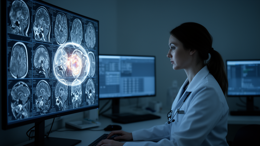
Every year, hundreds of thousands of people worldwide are diagnosed with meningioma, a tumor arising from the membranes that envelop the brain and spinal cord. It is the most common primary brain tumor in adults, accounting for roughly one in three cases. Yet beneath this familiar diagnosis lies a critical uncertainty: some meningiomas grow slowly and rarely return after removal, while others behave aggressively, invade surrounding tissue, and recur despite treatment. The only definitive way to tell them apart has long been to remove the tumor and examine it under a microscope. A new multicenter study, published in the Journal of Neuro-Oncology, suggests that artificial intelligence may begin to answer that question before a single incision is made, using nothing more than the MRI scans that patients already receive.
An international team led by researchers spanning institutions in Turkey and the United States assembled 455 histopathologically confirmed meningiomas from five tertiary referral centers, all operated between 2012 and 2025. Their goal was ambitious and methodologically rigorous: to predict, preoperatively, whether a tumor belonged to the World Health Organization’s Grade 1 category or the more dangerous Grades 2 and 3, and to determine which computational approach could do so reliably across different hospitals. The stakes are considerable. Accurate preoperative grading could reshape surgical planning, inform decisions about adjuvant radiotherapy, and calibrate how intensively patients are monitored after surgery.
The researchers built four fundamentally different prediction models, all drawing on contrast-enhanced T1-weighted MRI. The first was a classical radiomics model, which extracts thousands of handcrafted mathematical features describing tumor texture, shape, and intensity from both the tumor itself and a carefully constructed five-millimeter shell of surrounding brain tissue. The second harnessed nnU-Net, a self-configuring deep learning architecture famous in medical imaging circles, trained to simultaneously segment the tumor and encode its grade at the level of individual voxels. The third exploited DINOv3, a self-supervised vision transformer foundation model, using its frozen internal representations of tumor-centered image slices as input to a simple classifier. The fourth relied purely on clinical and conventional radiological variables: age, sex, tumor location, peritumoral edema, bone invasion, venous sinus invasion, and the dural tail sign.
What distinguishes this study from much of the AI-in-medicine literature is its uncompromising validation design. Models were developed on 346 patients from four centers using leave-one-center-out cross-validation, while a fifth center’s 109 patients were held out entirely, untouched until every model was frozen. All fifteen possible combinations of the four base models were prespecified and stacked with a linear meta-learner. No model was retrained, recalibrated, or discarded after external evaluation. The work followed the TRIPOD+AI and CLAIM reporting guidelines, and the authors deliberately avoided intensity harmonization between scanners, testing the models under realistic, messy, multicenter conditions rather than the curated environments that flatter many published algorithms.
The results delivered a striking verdict. In the development cohort, the radiomics model looked like the star, achieving an out-of-fold area under the curve of 0.800. But when transported to the unseen external hospital, its performance collapsed to 0.628, barely better than chance-adjacent guessing. Only two of its six carefully selected features retained statistical significance externally, and their effect sizes shrank dramatically. This pattern, in which handcrafted features capture site-specific scanner noise and spurious correlations rather than genuine biology, has haunted radiomics before, but rarely has it been demonstrated so cleanly within a single, leakage-free framework.
Deep learning told a different story. The nnU-Net segmentation-based classifier achieved the strongest external discrimination of any individual model, with an AUC of 0.732, followed closely by the DINOv3 transfer-learning model at 0.703. The clinical model managed 0.645. When the researchers stacked nnU-Net, DINOv3, and the clinical variables together, the multimodal ensemble reached the highest external AUC of the entire study, 0.742, significantly outperforming radiomics but not statistically distinguishable from nnU-Net alone. Within the ensemble’s linear meta-learner, nnU-Net contributed most to the final prediction, followed by DINOv3, while the clinical variables, chiefly peritumoral edema, bone invasion, and tumor location, played a smaller supporting role.
The study’s explainability analysis offers a glimpse of how such a system might function in practice. Because the meta-learner is linear and the clinical component decomposes into exact SHAP values, every individual prediction can be traced to its sources. In one external case, a WHO Grade 3 tumor received a high predicted probability driven overwhelmingly by concordant contributions from nnU-Net and DINOv3, with smaller pushes from bone invasion and edema. In another, a Grade 1 tumor’s low-risk prediction flowed from negative contributions by the same two deep learning components. This transparency matters: clinicians are far more likely to trust a probability they can interrogate than a black-box verdict, and the clinical variables, while adding little discriminative power, made the model’s reasoning legible.
Calibration and clinical utility analyses added further nuance. The ensemble’s external calibration slope of 1.32 indicated that its risk gradient survived transport, and decision-curve analysis showed net benefit over both treat-all and treat-none strategies at threshold probabilities of 0.23 and above. The apparent weakness of a 41 percent external sensitivity at the default threshold turned out to be largely an artifact of transferring a decision threshold between cohorts with different case mixes; when the threshold was set on development data to target 90 percent sensitivity, the model still caught 83 percent of high-grade tumors externally, albeit with specificity falling to 49 percent. The authors note that local recalibration of the intercept would be required before any clinical deployment, since class-balanced training skewed the predicted probabilities.
Perhaps the most sobering finding concerns the gap between development performance and real-world transportability. A development-only rule for choosing the best model would have selected a radiomics-containing combination with an external AUC as low as 0.685, a choice the external data exposed as misguided. The authors also acknowledge limitations: the external validation came from a single independent center, only one MRI sequence was used, Grade 3 tumors were too rare for separate analysis, molecular profiling was incomplete, and the reference standard spanned three editions of the WHO classification without central re-review. Conventional logistic regression using routine clinical variables performed surprisingly respectably externally, meaning the added value of learned image representations over a well-specified clinical model remains to be proven in larger cohorts.
Even so, the study marks a meaningful inflection point for AI in neuro-oncology. It demonstrates that learned deep learning representations, particularly those of nnU-Net, travel across institutions far better than handcrafted radiomic features, and it does so with a validation discipline that many earlier studies lacked. The external AUC of 0.742 is modest compared with the inflated figures of 0.90 and above often reported from single centers, but it may be a far more honest estimate of performance in heterogeneous, real-world practice. As the field moves toward imaging biomarkers that capture molecular risk and tumor biology beyond histological grade, this work offers both a template for rigorous evaluation and a clear warning: in medical AI, what looks brilliant in development may not survive the journey to the next hospital. Prospective validation in larger, geographically diverse cohorts is the necessary next step before these models can guide decisions in the neurosurgical operating room.
Subject of Research: MRI-based preoperative prediction of WHO grade in intracranial meningioma using deep learning, radiomics, and clinical models
Article Title: MRI-based preoperative WHO grade prediction in meningioma: development and comparison of deep learning, radiomics, clinical, and combined models in multicenter data
Article References: MRI-based preoperative WHO grade prediction in meningioma: development and comparison of deep learning, radiomics, clinical, and combined models in multicenter data. (n.d.). https://doi.org/10.1007/s11060-026-05827-6
Image Credits: AI Generated
DOI: 10.1007/s11060-026-05827-6
Keywords: meningioma, deep learning, radiomics, MRI, WHO grading, nnU-Net, DINOv3, external validation, machine learning, neuro-oncology, predictive modeling, brain tumor
News Source: Nathaniel Bowman. (October 14, 2026). AI Reads Brain Scans to Predict Tumor Aggressiveness Before Surgery. Scienmag.