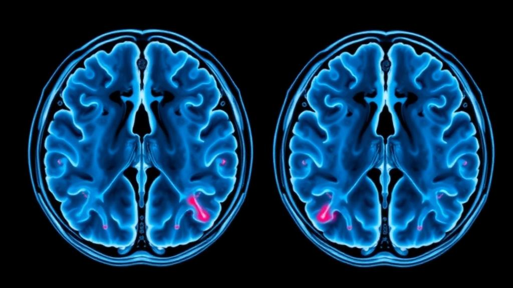
For decades, the gap between what radiologists see on an MRI scan and what pathologists observe under a microscope has been one of the quiet frustrations of neuro-oncology. Magnetic resonance imaging interrogates the brain at the scale of millimetres, while histopathology examines tissue at the scale of cells, and the two views have never been easy to reconcile. Now a team of researchers in China has built a framework that brings those two worlds into direct alignment, using surgical navigation technology to make sure that the tissue removed from a patient’s brain corresponds, point for point, to the exact spot imaged on the scanner. The result, published in the Journal of Translational Medicine, offers some of the strongest evidence yet that a sophisticated diffusion-based MRI technique can read out biologically meaningful information about the internal architecture of brain tumours.
The technique at the heart of the study is called continuous-time random walk, or CTRW, modelling. Standard diffusion-weighted MRI measures how freely water molecules move through tissue, which in turn reflects the density and organisation of cells. But conventional models assume that water diffusion behaves in a simple, orderly way. In reality, brain tumours are chaotic landscapes of dense cell clusters, necrotic debris, swollen oedema and tangled blood vessels, and water molecules navigating this terrain do not move in a simple pattern. CTRW mathematics abandons that assumption. It characterises diffusion using parameters that capture how far the movement departs from normal: the parameter β describes spatial heterogeneity, essentially how obstructed and irregular the microscopic environment is, while α describes temporal heterogeneity, capturing how the waiting times between molecular jumps deviate from ideal behaviour. A third parameter, the anomalous diffusion coefficient Dm, quantifies the overall magnitude of the anomalous diffusion process.
What makes the new study stand out is the extraordinary care taken to match imaging data with physical tissue. The researchers enrolled 98 adults undergoing brain tumour surgery at the Second Xiangya Hospital of Central South University, split across three diagnostic groups: 34 with high-grade gliomas, the most aggressive primary brain cancers; 34 with brain metastases, tumours that have spread from elsewhere in the body; and 30 with meningiomas, typically benign tumours arising from the brain’s protective membranes. During each operation, the surgical team used neuronavigation, the same GPS-like guidance system that helps surgeons locate tumours deep within the brain, to record the precise coordinates of a small specimen, roughly 2 millimetres across, taken from the superficial margin of the solid, contrast-enhancing part of the tumour.
That coordinate recording proved to be the crucial step. Back at the scanner, the team drew a matching 2-millimetre region of interest centred on the recorded coordinates, ensuring that the diffusion metrics extracted from the MRI data described the same sliver of tissue that the pathologist would later examine. Each specimen underwent haematoxylin and eosin staining, the workhorse technique of diagnostic pathology, along with immunofluorescence analysis capable of identifying specific immune cell populations. A pathologist scored the structural heterogeneity visible in each sample, and computational pathomics analysis added a quantitative layer to the assessment. By anchoring every imaging measurement to a precisely located tissue sample, the researchers eliminated the spatial guesswork that has undermined so many previous attempts to correlate MRI findings with pathology.
The findings are striking. In the malignant tumours, the spatial heterogeneity parameter β followed a clear gradient across brain regions: it was highest in the apparently healthy brain on the opposite side of the head, dropped in the peritumoral brain oedema that surrounds many tumours, and fell furthest within the contrast-enhancing tumour core itself. This makes intuitive sense once the underlying biology is considered. Healthy brain tissue, with its orderly arrangement of neurons and supporting cells, presents a relatively uniform environment for diffusing water. Oedema disrupts that order, and the densely packed, disorganised cells of an aggressive tumour disrupt it further. Most remarkably, β was inversely associated with the pathologist’s manual heterogeneity score with a Spearman correlation coefficient of −0.948, a near-perfect negative relationship that remained statistically significant after correction for multiple comparisons.
Beyond structure, the study reached into the tumour’s immune microenvironment. The researchers wanted to know whether the diffusion signature captured by CTRW could estimate the proportions of specific immune cells within the tumour, cells such as neutrophils, macrophages and B lymphocytes that play decisive roles in how brain tumours progress and respond to treatment. To test this, they compared five different regression model families using a rigorous validation scheme: repeated tumour-type-stratified nested cross-validation, a method that trains models on one subset of patients and tests them on patients the model has never seen, while keeping the tumour types balanced throughout.
Random-forest regression, a machine learning method that combines the predictions of many decision trees, emerged as the strongest performer. In a sensitivity analysis spanning 50 outer validation folds, the models estimated neutrophil proportions with a mean R² of 0.984, macrophage proportions with a mean R² of 0.976, and B cell proportions with a mean R² of 0.943. These are extraordinarily high figures for any imaging-based prediction, and the authors were careful to probe whether the models were simply learning to distinguish tumour types rather than reading genuine biology. When models were trained on tumour type alone, performance collapsed to mean R² values between 0.295 and 0.352. When the tumour-type signal was statistically removed from the training data, CTRW-based models still retained mean R² values of 0.717 for neutrophils, 0.767 for macrophages and 0.671 for B cells, indicating that the diffusion metrics carried real biological information beyond a simple tumour-type label.
The implications for clinical practice could be considerable. Brain tumours are notoriously heterogeneous, meaning that different regions of the same tumour can behave in radically different ways, harbour different genetic mutations and respond differently to therapy. Neurosurgeons and oncologists currently rely on contrast-enhanced MRI to define tumour boundaries, but that technique reveals only where the blood-brain barrier has broken down, not what the tissue is actually made of. A validated imaging biomarker that reports on structural disorganisation and immune cell composition without requiring a biopsy could help guide where surgeons take tissue samples, how radiation targets are drawn, and which patients might benefit from immunotherapies. Because the current study anchored its measurements to neuronavigation-recorded sampling sites, the framework could in principle be integrated directly into existing surgical workflows.
The authors are nonetheless emphatic about the limits of what they have shown. The associations they report reflect radiology-pathology correspondence at the sampling-site scale, not direct imaging of individual cells, and the immune-cell estimates, however impressive their internal validation, have not yet been tested as clinical predictors in prospective trials. It also remains to be established whether CTRW metrics add incremental value beyond the conventional diffusion measurements already available on standard clinical scanners. The study was approved by the institutional ethics committee and all participants gave written informed consent, and the work was supported by the National Natural Science Foundation of China and several Hunan Province research funds.
Even with those caveats, the study represents a methodological milestone. By fusing surgical navigation, advanced diffusion modelling and quantitative pathology into a single coherent pipeline, the researchers have demonstrated that the microscopic chaos of a brain tumour leaves a measurable fingerprint in the random walk of water molecules, one that a scanner can detect without opening the skull. If prospective studies confirm these findings, the humble diffusion MRI scan could evolve from a structural map into a molecular microscope, offering clinicians a non-invasive window into the biology of some of the most difficult tumours in human medicine.
Subject of Research: Continuous-time random walk diffusion MRI for characterising pathological heterogeneity in intracranial tumours
Article Title: Continuous-time random walk MRI and pathological heterogeneity in intracranial tumours: a neuronavigation-guided study
Article References: Gao, M., Zang, H., Yan, Y., Peng, J., He, Y., Liu, E., Zhao, W., Yuan, J., Lin, H., Liu, W., Zhou, Y., & Liu, J. (2026). Continuous-time random walk MRI and pathological heterogeneity in intracranial tumours: a neuronavigation-guided study. Journal of Translational Medicine. https://doi.org/10.1186/s12967-026-09072-3
Image Credits: AI Generated
DOI: 10.1186/s12967-026-09072-3
Keywords: brain tumours, continuous-time random walk, diffusion MRI, tumour heterogeneity, neuronavigation, high-grade glioma, brain metastasis, meningioma, immune cells, machine learning, radiology-pathology correlation, neuro-oncology
News Source: Nathaniel Bowman. (October 13, 2026). Random-Walk MRI Links Brain Tumour Scans to Microscopic Reality. Scienmag.