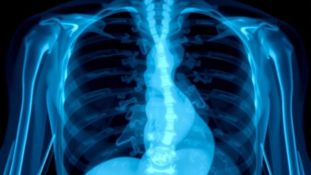
Artificial intelligence has promised to transform medical diagnosis for years, but the technology has always had an awkward dependency: it needs enormous amounts of high-quality data to learn from. During a pandemic, when AI-assisted diagnosis could matter most, collecting thousands of X-ray images from infected patients is slow, risky, and constrained by resources. A new study published in Cluster Computing by Walaa Saber, Ahmed El-Baz, and Rawya Rizk of Port Said University tackles this problem head-on with a generative model that can conjure realistic synthetic chest X-rays on demand—and, in the process, push diagnostic accuracy to 99.60 percent.
The framework, called Adaptive-CGAN, is a conditional generative adversarial network, a class of AI architecture in which two neural networks compete against each other. A generator produces synthetic images from random noise, while a discriminator tries to distinguish those fakes from real samples. As training proceeds, the generator becomes progressively better at fooling the discriminator, and the discriminator becomes sharper at catching deception. What makes Adaptive-CGAN distinctive is how it handles class information—the labels that tell the model whether an image depicts COVID-19, viral pneumonia, lung opacity, or a healthy chest. Rather than injecting the class label only once at the generator’s input, as conventional conditional GANs do, Adaptive-CGAN re-projects a learned class embedding at every layer of the generator, so that class information shapes both the global anatomical structure and the fine pathological texture of the output.
The second key innovation is a dual-output discriminator. Instead of merely judging whether an image is real or fake, the discriminator simultaneously classifies the image into its disease category. This means the generator receives two complementary streams of feedback during training: a realism signal that pressures it to suppress artifacts and produce anatomically plausible textures, and a class-alignment signal that forces generated images to carry the diagnostically relevant features of their target condition. The authors describe this as a dual-feedback mechanism, with both gradients flowing through a shared discriminator trunk back into the generator, aligning fine-scale, class-diagnostic cues without sacrificing visual plausibility.
To train and evaluate the model, the team compiled a multi-institutional chest X-ray dataset of roughly 8,000 unique images spanning four clinical classes, expanded through training-only augmentation to approximately 80,000 instances per epoch. The researchers took unusual care to prevent data leakage: duplicate and near-duplicate studies were removed before splitting, and partitioning into training, validation, and test sets was performed at the anonymized patient level, so no patient appeared in more than one subset. Augmentation was applied only after splitting and exclusively to the training data. Images were resized to 256 by 256 pixels, normalized, enhanced with histogram equalization, and denoised with median filtering before being paired with class labels encoded as 100-dimensional embedding vectors.
The results are striking. Against the closest baseline, AC-GAN, Adaptive-CGAN raised downstream classification accuracy from 93.75 percent to 99.60 percent, improved the Inception Score—a measure of generated image quality and diversity—by 17.65 percent, and reduced the Fréchet Inception Distance and Kernel Inception Distance, both standard measures of how closely synthetic images mimic real data distributions, by roughly 8 percent each. Training stability metrics reinforced the picture: conditional entropy fell from about 3.8 to 0.15 nats, the Jensen–Shannon divergence between real and generated distributions dropped to 0.041 compared with 0.119 for AC-GAN, and mode coverage reached 94.2 percent versus 63.2 percent for the baseline, indicating the model avoided the classic GAN failure mode of producing a narrow, repetitive set of outputs. An ablation study confirmed that both the layer-wise embedding and the dual-feedback discriminator contribute independently, and that combining them yields the largest gains.
Skeptics of generative medical imaging often worry about circularity: if you evaluate synthetic images with the same classifier used during training, the results may flatter the model. The authors addressed this with two independent validation strategies. A ResNet-50 network trained exclusively on real X-rays achieved 94.20 percent accuracy on unseen synthetic images, a modest 3.4-point degradation that suggests the synthetic data preserve diagnostically meaningful information. Separately, the team computed quality metrics in the feature space of a DenseNet-121 encoder pre-trained on the external CheXpert dataset of more than 224,000 chest X-rays, and Adaptive-CGAN outperformed AC-GAN in that independent, domain-specific representation as well.
The study also tackles a dimension that most generative modeling papers ignore: the environmental cost of training. Using the CodeCarbon tool, the researchers measured energy consumption and estimated CO₂ emissions with region-specific carbon-intensity factors. On identical local hardware, Adaptive-CGAN trained 15.8 percent faster and emitted 9.5 percent less CO₂ than AC-GAN, gains attributable to the architecture itself. Deployed on Google Cloud Platform, the model emitted just 0.0023 kilograms of CO₂, roughly half the 0.0047 kilograms of a local setup, thanks to optimized resource allocation and a TFRecords-based data pipeline that accelerates storage and loading. The cloud deployment also doubled throughput to 1 gigabit per second and cut latency from 100 milliseconds to 10, while the CWGAN baseline, by contrast, produced the highest emissions of any model tested at 0.0336 kilograms.
The cloud infrastructure is more than a convenience. The TFRecord pipeline serializes images into an efficient format for TensorFlow’s data-loading API, reducing input-output overhead and supporting scalable training on GPU-equipped virtual machines. The authors frame this as a distinct engineering contribution, arguing that as medical datasets grow toward multi-hospital and multi-national scale, optimized sharding, larger GPU memory, and distributed training will become essential. They also note that Google Cloud’s renewable energy commitments amplify the sustainability benefits, aligning the work with the United Nations Sustainable Development Goals on climate action and industrial innovation.
The researchers are careful to draw boundaries around what the technology can and cannot do. Generated images are synthetic and must never be treated as real patient cases or standalone diagnostic evidence; the intended use is research-level data augmentation and model development, not clinical decision-making. The team acknowledges remaining limitations, including potential artifacts, class ambiguity between visually similar thoracic conditions, the risk that biases in training data could be reproduced or amplified, and the possibility of memorization, which they probed with nearest-neighbor inspection. They call for prospective clinical validation, blinded specialist review, privacy safeguards such as differential privacy and membership-inference testing, and fairness audits across patient subgroups before any clinical-facing deployment.
Even within those limits, the work signals where medical AI is heading. Generative models that can produce faithful, class-controlled synthetic images could relieve the data bottleneck that has long constrained diagnostic deep learning, particularly for rare diseases and fast-moving epidemics. By demonstrating that architectural innovation, rigorous validation, cloud-scale engineering, and carbon accounting can be pursued together, the Port Said team offers a template for what sustainable, clinically responsible generative AI might look like as the field matures.
Subject of Research: A conditional generative adversarial network for synthesizing medical chest X-ray images on cloud infrastructure to improve disease diagnosis and reduce carbon emissions
Article Title: Adaptive-CGAN: a comprehensive framework for generative modeling on google cloud platform
Article References: Saber, W., El-Baz, A., & Rizk, R. (2026). Adaptive-CGAN: a comprehensive framework for generative modeling on google cloud platform. Cluster Computing, 29(12), Article 710. https://doi.org/10.1007/s10586-026-06496-w
Image Credits: AI Generated
DOI: 10.1007/s10586-026-06496-w
Keywords: generative adversarial networks, medical imaging, synthetic data, COVID-19, chest X-ray, Google Cloud Platform, deep learning, diagnostic accuracy, sustainable AI, carbon emissions, data augmentation, TFRecords
News Source: Ophelia Keating. (October 13, 2026). AI Learns to Fake Chest X-Rays So Well It Boosts Disease Diagnosis to Near Perfection. Scienmag.