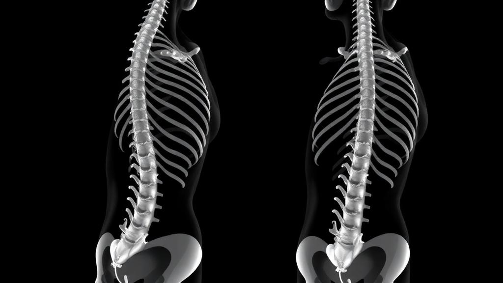
Artificial intelligence has transformed medical imaging, but it has a blind spot that surgeons know all too well: the severely crooked spine. Deep learning models that slice through ordinary CT scans with near-perfect accuracy tend to falter when confronted with the twisted, rotated vertebrae of severe scoliosis, precisely the cases where surgical planning demands the most reliable anatomy maps. A team at the University of Basel and University Children’s Hospital Basel now reports a strikingly simple fix. Instead of hunting for rare, painstakingly annotated scans of deformed spines, they taught their algorithms to learn from the unseen by bending healthy spines into scoliosis-like shapes, generating synthetic training data that dramatically improved segmentation of real, severely deformed patients.
The problem the researchers set out to solve is fundamentally one of scarcity. Scoliosis, a three-dimensional deformity of the spine quantified by Cobb angles, can progress rapidly in adolescents, and surgical correction is typically considered when the curve reaches 45 to 50 degrees or when documented progression and symptoms appear. Modern correction relies on posterior spinal fusion with pedicle screws and bilateral rods, procedures that are technically demanding and increasingly performed with computer assistance. Preoperative planning of screw trajectories and rod contouring, 3D-printed rehearsal models, and augmented-reality navigation systems all depend on accurate 3D vertebral segmentation derived from imaging. Yet the deep learning models behind this segmentation are data-hungry, and datasets of annotated severe scoliosis cases are scarce and, to the authors’ knowledge, not publicly available.
Conventional data augmentation was never designed to close this gap. Standard schemes perturb images locally, adding noise, adjusting intensity, cropping, or applying mild elastic deformations, but they do not reproduce the global, pathology-specific shape changes that define severe scoliosis. The Basel team, led by Volodimir Buchakchiyskiy and Philippe C. Cattin, therefore built a pathology-informed, anatomy-aware pipeline that synthesizes scoliosis-like deformations by jointly warping CT images and their segmentation masks, using only publicly available data from the VerSe 2019 and 2020 MICCAI challenge datasets. After merging and deduplicating, they obtained 408 source volumes of anatomically normal spines, from which they generated synthetic variants offline, with no manual review or filtering.
The technical heart of the method is an elegant geometric construction. The pipeline begins by extracting vertebral centroids from a segmentation mask and fitting a smooth cubic spline through them to define the spinal centerline. This healthy centerline is then deformed into a randomized pathological shape using a fourth-degree Bézier curve, a choice that offers enough flexibility to reproduce both C-shaped and S-shaped curvatures typical of scoliosis while naturally limiting unrealistic higher-order oscillations. Three intermediate control points are randomly displaced within bounded offsets and refined to maximize curvature while preserving arc length, and the optimized curve is reparametrized as a cubic spline in the same basis as the original.
Extending this curve-based deformation into the full 3D volume is where the method becomes genuinely sophisticated. The researchers construct local orthonormal coordinate frames along both the healthy and deformed centerlines using parallel transport, then define circular polar grids of control points within each frame, with the grid radius set adaptively from the median vertebral bounding box. When the deformed spline bends sharply, neighboring control planes risk overlapping or crossing, so the team enforces an order-preserving, arc-length monotonic mapping that guarantees no point moves backward along the curve, preventing fold-overs in the deformation field. The sparse correspondences are then interpolated into a continuous volumetric backward map via a piecewise-linear tetrahedral mesh built by 3D Delaunay triangulation, with barycentric weights within each tetrahedron yielding the undeformed coordinate of any point. Applying this map to the original CT volume, with linear interpolation for intensities and nearest-neighbor interpolation for masks, yields anatomically consistent synthetic scans that preserve vertebral topology while exhibiting artificial scoliosis-like deformation.
To test whether this synthetic diversity actually helps, the team trained sixteen models under strictly matched conditions: the standard 3D full-resolution nnU-Net v2 convolutional network and the transformer-based nnU-Net v2 PRIMUS, each trained on four different training sets with two augmentation regimes, all on an NVIDIA V100 with a fixed budget of 250,000 iterations and 1,000 epochs. Evaluation was performed on an in-house cohort of 18 adolescent scoliosis CT volumes from a pediatric hospital, covering variable severity and etiology, and on the 1,005-scan public Spine1K dataset as a large benchmark of predominantly non-scoliotic anatomy. The results were unambiguous. The best convolutional network setting, trained on the original VerSe scans plus a doubled set of synthetic scans, increased mean Dice from 0.765 to 0.842 and the vertebra success rate, the percentage of vertebrae with Dice at or above 0.8, from 73.2 to 81.6 percent, while reducing the 95th percentile of the 95-percentile Hausdorff distance from 31.1 to 24.9 millimeters.
The gains were largest exactly where it matters most for surgery. In an apex-focused analysis using 55 surgeon-verified vertebral labels at the curve apex, the most disrupted and technically challenging region, mean Dice jumped from 0.720 to 0.854, the vertebra success rate rose from 65.5 to 81.8 percent, and the HD95 tail dropped from 54.1 to 22.4 millimeters. A severity analysis across 17 cases with Cobb angle measurements, grouped into low, medium, and high curvature clusters, showed that the benefit increases with deformity: the high-curvature group, with a mean Cobb angle of 105 degrees, achieved a statistically significant mean Dice gain of 0.114, while the medium-curvature group at 62.1 degrees gained 0.064 and approached significance. Crucially, adding only generic elastic deformations to the standard pipeline improved the baseline far less, from 0.765 to 0.794, confirming that the advantage comes from modeling global scoliosis-like curvature rather than local shape variability.
Just as important is what the synthetic data did not do. On Spine1K, mixed real-synthetic training preserved high performance, with the best convolutional mean Dice of 0.908 compared with 0.904 for the real-data baseline, and the transformer improving from 0.886 to 0.903. But training on synthetic data alone was less reliable, particularly for the transformer, producing worse HD95 tails and unstable results. The message is clear: synthetic scoliotic scans should supplement real training data, not replace them. Doubling the synthetic data proved the sweet spot, while quadrupling it brought diminishing returns, possibly reflecting a distribution mismatch between synthetic and real anatomy at higher synthetic ratios.
The clinical implications extend beyond a leaderboard metric. More reliable segmentation in severe scoliosis reduces the manual correction effort clinicians must invest and improves the robustness of downstream tasks such as vertebral labeling and measurement. The higher vertebra success rate indicates fewer catastrophic failures in exactly the challenging anatomy where clinical pipelines typically require the most human intervention. Because the entire synthetic generation and post-processing pipeline is fully automated, requiring no manual inspection or curation, the approach could in principle be applied wherever annotated pathological data are scarce but healthy anatomy is abundant, a situation that describes most rare and severe conditions in medicine.
The authors are candid about limitations. The test cohort is small and single-institution, with 18 cases and Cobb angles available for 17; warping and resampling can introduce interpolation artifacts and local distortions; and the elastic-deformation baseline was tuned only once on a single fold before being frozen. The severity analysis was exploratory and awaits validation on broader multi-center cohorts across scanners and protocols. Still, the core finding stands: anatomy-aware synthetic deformations, generated from public data alone and without any new annotations or architecture changes, can meaningfully augment spine datasets and improve robustness under clinically realistic distribution shifts. For a field where the rarest patients are often the ones who need algorithms most, learning from the unseen may prove one of the most practical ideas in medical AI this year.
Subject of Research: Synthetic data augmentation for AI-based 3D vertebral segmentation in severe scoliosis CT imaging
Article Title: Learning from the unseen: synthetic data for scoliosis AI segmentation
Article References: Buchakchiyskiy, V., El Hadramy, S., Zentai, N., Wehrli, M., Krol, Z., Cescato, G., Licci, M., Studer, D., Hasler, C. C., & Cattin, P. C. (2026). Learning from the unseen: synthetic data for scoliosis AI segmentation. International Journal of Computer Assisted Radiology and Surgery. https://doi.org/10.1007/s11548-026-03782-1
Image Credits: AI Generated
DOI: 10.1007/s11548-026-03782-1
Keywords: scoliosis, synthetic data, data augmentation, CT segmentation, deep learning, nnU-Net, vertebral segmentation, spine surgery, medical imaging, Bézier deformation, VerSe dataset, Spine1K
News Source: Ophelia Keating. (October 12, 2026). Fake spines, real gains: synthetic CT data teach AI to segment severe scoliosis. Scienmag.