Brain tumors remain one of the most feared diagnoses in medicine, and the first line of defense is often a radiologist peering at magnetic resonance imaging scans. That first look matters enormously: delayed or inaccurate interpretation can change the entire course of treatment. Yet manual reading of MRI scans is slow, and studies have long shown that different specialists can disagree about what they see in the same image. A new study published in Cluster Computing by Md. Alamin Talukder of the International University of Business Agriculture and Technology in Dhaka and Majdi Khalid of Umm Al-Qura University in Saudi Arabia tackles this problem head-on with an artificial intelligence framework that not only classifies brain tumors with accuracy approaching 100 percent, but also shows clinicians exactly where it looked.
The framework, called XIM+CMAES, is built on an idea that has become central to modern medical AI: ensemble learning. Rather than trusting a single neural network, the system combines four well-known transfer learning models—Xception, InceptionV3, MobileNet, and MobileNetV2. Each of these networks was originally trained on millions of natural images, then fine-tuned for the task of spotting tumors in brain scans. The four architectures bring different strengths to the table. Xception, with its depthwise separable convolutions, excels at capturing fine-grained spatial patterns; InceptionV3 processes features at multiple scales simultaneously; and the two MobileNet variants are lightweight models designed for computational efficiency, making them attractive for deployment in clinics with limited hardware.
The genuinely novel element is how the ensemble is fused. In a typical ensemble, the outputs of member models are averaged or combined with fixed weights. Here, the researchers turned to the Covariance Matrix Adaptation Evolution Strategy, or CMAES, a powerful derivative-free optimization algorithm borrowed from evolutionary computation. CMAES searches for the optimal set of weights to assign to each model’s contribution by iteratively sampling candidate solutions from a multivariate probability distribution, then adapting the covariance matrix of that distribution based on which candidates perform best. This allows the optimizer to learn correlations between the models’ errors and navigate the high-dimensional weight space efficiently, without ever computing gradients. The result is an adaptive fusion that exploits the complementary strengths of the four networks and stabilizes predictions across diverse imaging conditions.
The performance figures reported in the study are striking. On the BTMRI dataset, XIM+CMAES achieved 99.79 percent accuracy and 99.93 percent specificity. On BTCMRI, a harder multi-class benchmark, it reached 98.16 percent accuracy with 99.38 percent specificity. On the SIAR dataset, it posted 99.93 percent accuracy and 99.92 percent specificity. Crucially, the ensemble consistently outperformed each of the individual transfer learning baselines it was built from, demonstrating that the evolutionary fusion was doing real work rather than merely averaging away noise. Specificity deserves particular attention in a clinical context: a highly specific model rarely raises false alarms, which means fewer patients sent into unnecessary anxiety and unnecessary follow-up procedures.
Generalization has long been the Achilles heel of deep learning in medical imaging. A model trained on one hospital’s scanner and patient population often falters when confronted with images from another. By evaluating on three publicly available, heterogeneous MRI datasets—each with different acquisition parameters, class distributions, and tumor subtypes—the authors directly targeted this weakness. The consistent near-ceiling performance across all three suggests the framework is not simply memorizing the quirks of a single dataset, a failure mode that has undermined many earlier claims of superhuman diagnostic AI.
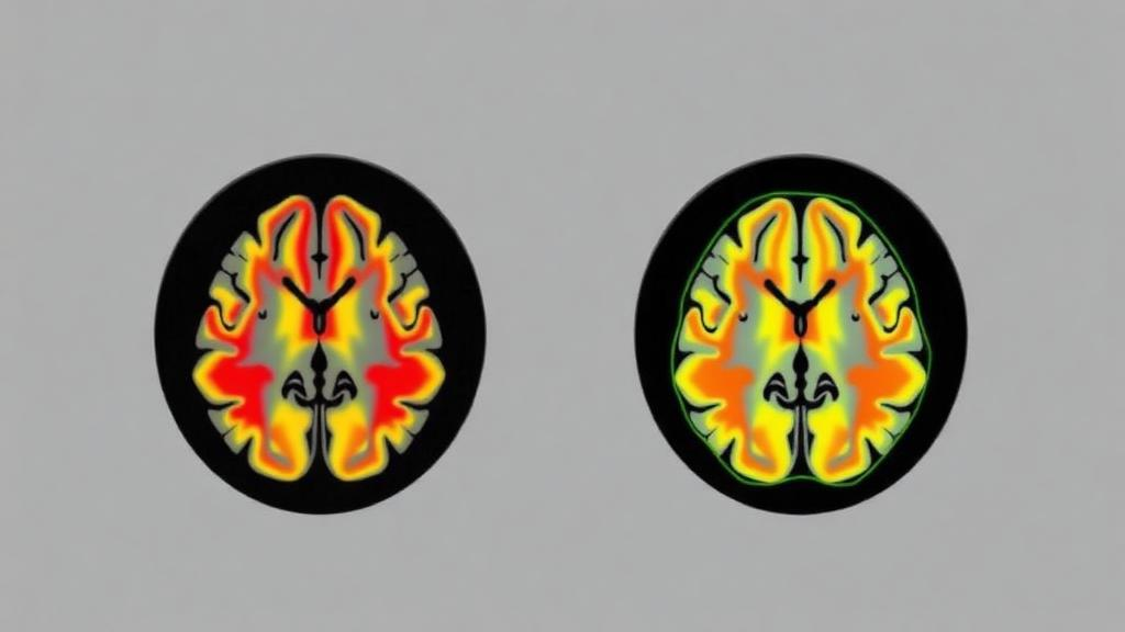
But raw accuracy alone has never been enough to bring deep learning into the clinic. Radiologists and regulators alike have grown wary of black-box models that issue verdicts without justification, especially in a domain as consequential as oncology. To address this, the researchers embedded explainable AI techniques into the framework, including Weighted Grad-CAM, SHAP, and LIME. Grad-CAM and its weighted variant generate heatmaps that highlight the image regions most responsible for the model’s decision, effectively letting the network point at the suspicious tissue. SHAP, based on game-theoretic Shapley values, quantifies how much each feature contributed to a given prediction, while LIME builds a simple local surrogate model around each individual prediction to explain it in understandable terms.
The practical effect of these explanations is to turn the AI from an oracle into a collaborator. A radiologist reviewing the system’s output can see whether the highlighted regions correspond to the tumor’s actual location and appearance, and can discount the prediction if the model appears to be latching onto irrelevant artifacts such as scanner noise or anatomical landmarks. This kind of visual verification supports radiological decision-making rather than replacing it, and it aligns with the broader movement toward precision oncology, where treatment decisions are increasingly tailored to the specific characteristics of each patient’s disease.
The study also situates itself within a crowded and fast-moving field. The reference list catalogs a wave of recent work applying transfer learning, vision transformers, genetic algorithms, particle swarm optimization, Bayesian optimization, and hybrid CNN-transformer architectures to brain tumor classification. Many of these approaches optimize hyperparameters or feature extraction with metaheuristics, but the combination of an Xception-Inception-MobileNet ensemble with CMAES-driven weight optimization, paired with a multi-method explainability layer, appears to be a distinctive configuration. The authors report that the framework is computationally efficient as well, an important consideration given that the MobileNet backbone models were explicitly designed for resource-constrained environments.
There are, as always, caveats worth keeping in mind. The evaluation relied on publicly available Kaggle datasets rather than prospective clinical trials, and real-world deployment would require validation on freshly acquired scans from diverse clinical sites, along with regulatory review. The authors note that the study involved no human participants or animals and required no ethical approval, and they declare no competing interests and no external funding. Still, the trajectory of the work is clear: automated diagnostic tools are moving from laboratory curiosities toward genuine clinical decision support, and the combination of evolutionary optimization and explainability represents one of the more thoughtful paths forward.
If the reported results hold up under clinical scrutiny, the implications could be significant for global health. Brain tumors impose a heavy burden worldwide, and access to expert neuroradiologists is unevenly distributed, particularly in low- and middle-income countries where pediatric and adult brain tumor care often suffers from delayed diagnosis. A lightweight, accurate, and transparent AI assistant that runs on modest hardware could help close that gap, flagging urgent cases for expert review and giving clinicians in under-resourced settings a second opinion that arrives in seconds. The XIM+CMAES framework is a research result, not yet a bedside tool, but it offers a compelling preview of how evolution-inspired algorithms and explainable AI may soon work together to make one of medicine’s most difficult visual judgments faster, more consistent, and more trustworthy.
Subject of Research: Explainable ensemble deep learning with CMAES optimization for brain tumor classification from MRI images
Article Title: An explainable ensemble deep learning framework for brain tumor classification using covariance matrix adaptation evolution strategy
Article References: Talukder, M. A., & Khalid, M. (2026). An explainable ensemble deep learning framework for brain tumor classification using covariance matrix adaptation evolution strategy. Cluster Computing, 29(13), Article 735. https://doi.org/10.1007/s10586-026-06452-8
Image Credits: AI Generated
DOI: 10.1007/s10586-026-06452-8
Keywords: brain tumor classification, deep learning, ensemble learning, CMAES optimization, transfer learning, MRI, explainable AI, SHAP, LIME, Grad-CAM, precision oncology, clinical decision support
News Source: Cassandra Pierce. (October 10, 2026). Evolution-Tuned AI Ensemble Reads Brain Tumor Scans With Near-Perfect Accuracy. Scienmag.