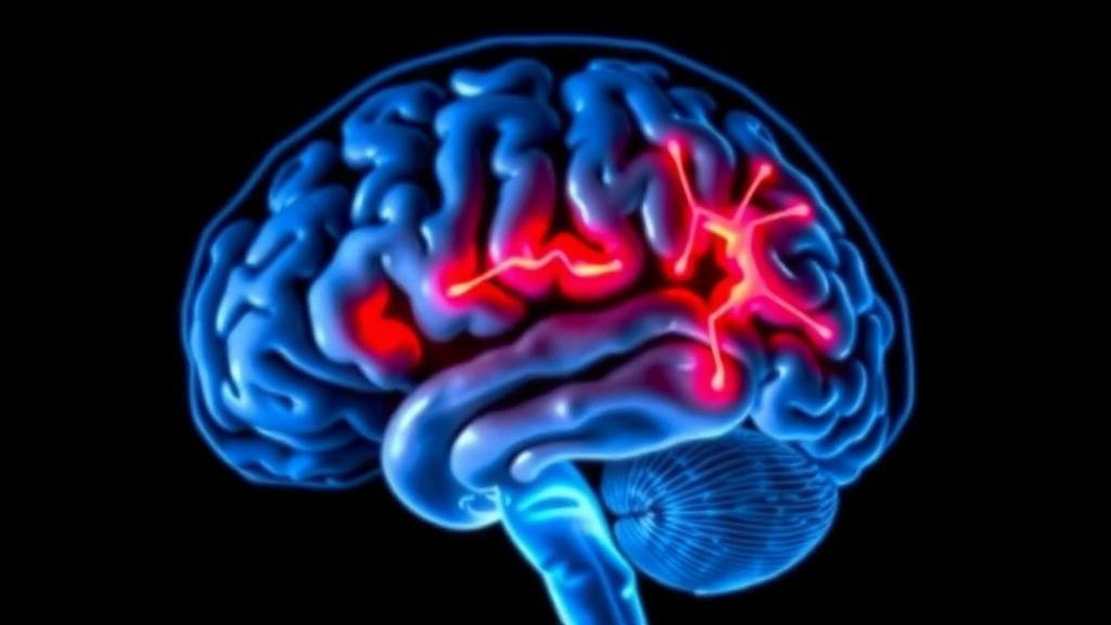
Parkinson’s disease has long been diagnosed by what it does to the body: the tremor, the stiffness, the shuffling gait that announce its arrival only after the brain has already suffered years of silent damage. But a new study published in npj Parkinson’s Disease suggests that the disease leaves detectable fingerprints on brain function far earlier than anyone can see them in a clinic. Using a sophisticated form of magnetic resonance imaging and machine learning, an international team of researchers has shown that the characteristic functional network disruptions of Parkinson’s disease can be identified not only in patients with early symptoms, but also in people who carry genetic risk factors yet remain entirely symptom-free.
The research, led by Amgad Droby of Tel Aviv Sourasky Medical Center together with colleagues at the Feinstein Institutes for Medical Research in New York and collaborators at several other institutions, focused on a question that has haunted the field of neurodegeneration for decades: can we see Parkinson’s disease before it becomes Parkinson’s disease? The answer, according to this work, appears to be a qualified but encouraging yes. The team analyzed 295 resting-state functional MRI datasets drawn from 52 people with early-stage Parkinson’s disease, 92 first-degree relatives who carry Parkinson’s-associated mutations in the GBA1 or LRRK2 genes but show no symptoms, and 58 healthy non-carriers who served as controls.
The technical heart of the study lies in how the researchers processed those scans. Resting-state functional MRI measures spontaneous fluctuations in blood oxygenation across the brain while a person lies still, doing nothing in particular. These fluctuations reveal the brain’s intrinsic functional architecture: the networks of regions that rise and fall in activity together. Rather than examining individual connections one by one, the team used independent component analysis, a mathematical technique that decomposes the massive four-dimensional imaging data into spatial patterns representing distinct functional networks. This approach allows researchers to capture whole-network topographies, the characteristic spatial layouts of brain activity, rather than isolated pairwise links.
What makes this study particularly clever is its borrowing from a different imaging modality. For years, researchers studying Parkinson’s have relied on positron emission tomography with the fluorodeoxyglucose tracer to identify two reproducible metabolic patterns: the Parkinson’s disease-related pattern, known as PDRP, which is elevated in the motor phase of the illness, and the cognition-related pattern, or PDCP, which tracks cognitive decline. These patterns have proven valuable for diagnosis and for measuring disease progression in clinical trials, but PET imaging involves radiation exposure and is expensive and logistically demanding. The central question of the new work was whether equivalent patterns could be extracted from radiation-free MRI scans, and whether they would behave the same way in people who do not yet have symptoms.
To find out, the researchers combined the independent component analysis with a support vector machine, a classical machine learning classifier trained to distinguish the brain patterns of Parkinson’s patients from those of healthy non-carriers. The classifier achieved 87 percent accuracy in that primary discrimination task, with an average accuracy of 78 percent across the full set of group comparisons. Once the classifier had learned to separate the groups, the team applied local interpretable model-agnostic explanations, a technique designed to open the black box of machine learning and reveal which features of the data drove each decision. This step isolated two fMRI-derived patterns that the investigators named fPDRP and fPDCP, the functional analogues of the well-known PET patterns.
With these patterns in hand, the researchers computed expression scores for every participant, essentially asking how strongly each individual’s brain expressed the Parkinson’s-related and cognition-related network topographies. The results were striking. Patients with early Parkinson’s disease showed significantly higher expression of both fPDRP and fPDCP than any of the preclinical groups. But within the preclinical population, an important divergence emerged. Participants classified as having a high likelihood risk, defined as a score of 60 or above on the Movement Disorder Society’s criteria for prodromal Parkinson’s, showed elevated fPDRP expression compared with lower-risk carriers, while their fPDCP expression remained unchanged. In other words, the motor-related network signature appears to rise first, before the cognitive signature, mirroring the clinical course of the disease itself.
Perhaps the most clinically consequential finding concerns what did not matter. Neither the specific genetic mutation a carrier carried, whether GBA1 or LRRK2, nor the status of their cerebrospinal fluid alpha-synuclein seed amplification assay, a molecular test that detects misfolded alpha-synuclein protein, significantly affected network expression in people who already had clinical Parkinson’s disease. This suggests that once the disease manifests, the functional network disruption converges on a common topography regardless of the underlying biology that triggered it. The network patterns, in this sense, capture the final common pathway of the disease rather than its upstream causes.
The team did not stop at cross-sectional analysis. To test whether the fMRI-derived patterns were stable and meaningful over time, they validated their findings in an independent cohort of 33 non-manifesting carriers who had been followed for ten years as part of a long-term observational study. The patterns proved reproducible in this validation cohort, lending weight to the idea that they represent genuine biological signals rather than statistical artifacts. Among the validation participants, eight individuals converted to manifest Parkinson’s disease during the follow-up period. Those converters showed numerically higher baseline fPDRP expression than those who did not convert, though the difference did not reach statistical significance, likely because of the small number of converters. The direction of the effect, however, is exactly what one would hope to see in a genuine preclinical marker.
The implications of this work extend well beyond the laboratory. Drug development for Parkinson’s disease has been repeatedly frustrated by the difficulty of testing therapies in people whose brains are already substantially damaged by the time of diagnosis. A reliable, radiation-free marker of preclinical network disruption could allow trials to enroll people at the earliest stages of the disease process, when interventions have the best chance of altering its trajectory. Because resting-state fMRI involves no ionizing radiation, it could in principle be repeated frequently, enabling researchers to track network changes over time in a way that PET-based measures cannot easily support. The study was supported by Biogen and by the Michael J. Fox Foundation for Parkinson’s Research, both of which have invested heavily in the search for reliable biomarkers of early disease.
Cautions remain, of course. The high-risk group in the preclinical analysis was defined by clinical criteria rather than by certain future diagnosis, and the conversion analysis, while directionally encouraging, involved only eight individuals. The authors themselves note that the fPDRP differences between converters and non-converters were not statistically significant. Still, the convergence of evidence across independent cohorts, multiple genetic risk groups, molecular CSF testing, and a decade of longitudinal follow-up makes a compelling case that the brain’s functional network architecture carries readable information about Parkinson’s disease risk long before the first tremor. For the millions of people who carry Parkinson’s-associated mutations and live with the uncertainty of what the future holds, the prospect of seeing the disease coming, and perhaps one day stopping it before it starts, has moved measurably closer.
Subject of Research: Resting-state fMRI detection of Parkinson's disease-related functional network patterns in preclinical and early disease stages
Article Title: Resting state fMRI network topographies in preclinical and early Parkinson’s disease stages
Article References: Droby, A., Nguyen, N., Do, P., Truong, J., Marebwa, B., Cedarbaum, J. M., Mirelman, A., Eidelberg, D., Vo, A., & Thaler, A. (2026). Resting state fMRI network topographies in preclinical and early Parkinson’s disease stages. npj Parkinson's Disease. https://doi.org/10.1038/s41531-026-01584-5
Image Credits: AI Generated
DOI: 10.1038/s41531-026-01584-5
Keywords: Parkinson's disease, resting-state fMRI, biomarkers, GBA1, LRRK2, machine learning, independent component analysis, PDRP, PDCP, alpha-synuclein, prodromal Parkinson's, neuroimaging
News Source: Cassandra Pierce. (October 10, 2026). Brain Scan Fingerprints of Parkinson’s Emerge Years Before Symptoms Appear. Scienmag.