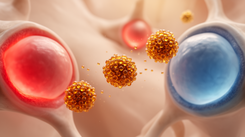
Osteoarthritis has long been dismissed as simple wear and tear of cartilage, but a growing body of evidence paints a very different picture: a chronic, self-perpetuating inflammatory disease in which the joint’s own lining becomes a factory for destructive signals. Now, a team of researchers writing in Advanced Science has unveiled an ingeniously targeted nanomedicine that attacks the disease at its metabolic roots. Their mannose-modified selenium nanoparticles, injected directly into the joint, home in on the most inflamed cells in the synovium, restore the tissue’s failing antioxidant defenses, and throttle the runaway sugar metabolism that keeps the inflammation burning. In mice, the treatment eased pain-like behaviors, improved gait, and slowed cartilage degeneration over twelve weeks of therapy.
The story begins with a puzzle the researchers uncovered by dissecting the synovium, the soft tissue lining the joint capsule, at single-cell resolution. Using single-cell RNA sequencing of human osteoarthritic tissue, they identified seven major cell populations and found that two of them—fibroblast-like synoviocytes, the structural workhorses of the synovium, and infiltrating macrophages—stood out with dramatically elevated glycolytic activity. These cells cranked up expression of SLC2A1, the gene encoding the glucose transporter GLUT1, along with PFKFB3, a key enzyme that amplifies glycolytic flux. Immunofluorescence staining of human knee synovium and of mouse models confirmed that GLUT1 accumulated precisely in the activated, tumor-necrosis-factor-producing cells driving the inflammation. Pseudotime analysis suggested that high SLC2A1 expression marked a terminal, pathologically transformed cell state, and the GLUT1-high subsets carried strong inflammatory, fibrotic, and catabolic signatures.
But the metabolic story had a second, darker thread. Excessive glycolysis is tightly linked to oxidative stress, and the team found that the synovium’s selenium-dependent antioxidant system was in disarray. Mass spectrometry revealed reduced selenium levels in the synovial fluid of osteoarthritis patients, and single-cell analysis showed broad dysregulation of selenoprotein genes, including the antioxidant regulators TXNRD1 and SELENOH, across multiple cell populations. Strikingly, the GLUT1-high cells that were burning glucose fastest also showed the lowest selenoprotein expression and the highest oxidative stress scores, an inverse relationship confirmed by spatial analysis of tissue sections. The picture that emerged was a vicious cycle: glucose flooding in through GLUT1 feeds inflammatory signaling, inflammatory signaling suppresses the selenoprotein shield, and the resulting reactive oxygen species further entrench the inflammatory state.
Selenium’s role here is not incidental. The trace element is incorporated into selenoproteins as the amino acid selenocysteine, which sits at the catalytic heart of enzymes that neutralize peroxides and maintain cellular redox balance. Selenium deficiency is a recognized cause of joint disease in humans—Kashin-Beck disease, an endemic osteoarthropathy, is the classic clinical archetype—and prior work has shown that selenium supplementation protects cartilage. What remained unclear was how selenium status affected the inflamed synovium itself, and whether it could be exploited therapeutically. The researchers’ answer was to build a delivery vehicle that would exploit the very metabolic quirk that makes osteoarthritic synoviocytes pathological: their GLUT1-overexpressing, glucose-hungry surface.
The vehicle they engineered is elegantly simple in concept. Spherical selenium nanoparticles roughly 100 nanometers in diameter were synthesized by reducing sodium selenite with ascorbic acid in the presence of a PEGylated lipid, then decorated with mannose, a sugar that hijacks GLUT1-mediated uptake pathways. Transmission electron microscopy and dynamic light scattering confirmed uniform spheres with excellent colloidal stability across seven days at lysosomal, inflammatory, and physiological pH values. Fourier-transform infrared spectroscopy verified the carbohydrate coating through its characteristic O-H, C-H, and C-O stretching vibrations, and the zeta potential shifted from −2.7 to −1.2 millivolts after modification, a fingerprint of successful grafting. Cytotoxicity testing in synoviocytes and macrophages showed viability above 95 percent at concentrations up to 20 micromolar over 24 hours, setting a safe working dose well within international biocompatibility standards.
What happens to the nanoparticles once they enter a cell proved just as important as getting them there. Using high-performance liquid chromatography coupled with inductively coupled plasma mass spectrometry, the team tracked the intracellular fate of the selenium and found that the nanoparticles were rapidly metabolized into bioactive organic species, predominantly selenocystine and methylselenocysteine, the very precursors cells use to build selenoproteins. In other words, the particles do not merely dump an inert antioxidant into the cell; they feed the cell’s own selenoprotein assembly line. Quantitative proteomics of inflamed synoviocytes treated with the particles bore this out: of 8,868 proteins quantified, the antioxidant selenoprotein TXNRD1 more than doubled, and several other redox- and endoplasmic-reticulum-associated selenoproteins rose alongside it, while inflammatory mediators such as IL-6, CXCL1, CCL2, VCAM-1, and ICAM-1 fell.
The metabolic reprogramming was equally striking. Functional assays using Seahorse flux analysis showed that treated cells reduced their extracellular acidification rate, glucose uptake, and lactate production—hallmarks of glycolysis—while increasing their oxygen consumption rate, a sign that mitochondrial respiration was being restored. The particles suppressed the pro-glycolytic enzyme PFKFB3 and raised TXNIP, a known brake on glucose uptake, and the transcription factor ETS1, which correlates strongly with PFKFB3 in osteoarthritic tissue, declined in parallel. Mitochondrial membrane potential, which collapses under inflammatory oxidative assault, was stabilized as measured by JC-1 staining. In macrophages, the treatment pushed polarization away from the pro-inflammatory M1 phenotype, reducing expression of Il1b, Il6, Tnfa, Nos2, and Cd86, and shifting the population toward reparative M2-like cells in the mouse synovium.
Delivery was the make-or-break question for any intra-articular therapy, and here the mannose modification paid visible dividends. In a surgically induced mouse model of osteoarthritis, fluorescently labeled mannose-coated particles persisted in the knee joint for roughly fourteen days after a single injection, compared with about ten days for unmodified particles. Using genetically engineered reporter mice in which fibroblast-like synoviocytes glow green and myeloid cells glow red, the researchers showed that the modified particles accumulated preferentially inside both target cell types in the inflamed synovium. Systemic distribution was limited, with the liver as the main clearance organ, and histological, blood-count, and serum chemistry analyses found no signs of organ injury or toxicity after repeated dosing.
The functional payoff was measurable in the animals’ own behavior. Mice treated with the nanoparticles every two weeks for twelve weeks showed higher paw withdrawal thresholds on von Frey testing, meaning less mechanical hypersensitivity, along with longer rotarod latency and normalized gait parameters including stride length, swing time, and weight-bearing symmetry between the operated and healthy limbs. Expression of TRPA1, a ROS-sensing ion channel in sensory neurons and synoviocytes that has been implicated in osteoarthritic pain, was suppressed both in the dorsal root ganglia and in the joint lining. Histology told the same story from the inside: reduced synovial lining thickening and inflammatory infiltration, preserved proteoglycan content in the cartilage, lower OARSI degeneration scores, and diminished MMP13, the matrix-degrading enzyme that serves as a barometer of both cartilage catabolism and synovial inflammation.
The authors are careful about what their data do and do not prove. The restoration of TXNRD1 accompanied the anti-inflammatory effects but has not yet been shown to be causally required, and the ETS1–PFKFB3 relationship remains an association pending direct regulatory validation. Whether TRPA1 suppression is a direct effect of the selenium or a secondary consequence of a calmer joint environment is also unresolved. Even so, the study delivers a compelling proof of concept: a disease-modifying osteoarthritis therapy that does not simply block a single cytokine, but instead rebuilds the metabolic and redox architecture of the inflamed synovium from within. By turning the GLUT1-high phenotype of diseased synoviocytes from an accomplice of pathology into a delivery address, the work opens a genuinely new avenue for treating a condition that afflicts hundreds of millions of people worldwide and currently has no cure.
Subject of Research: Mannose-modified selenium nanoparticles that remodel glycolysis and redox homeostasis in inflamed synovium for osteoarthritis therapy
Article Title: Metabolic‐Redox Remodeling of Inflammatory Synovium by Mannose‐Modified Selenium Nanoparticles for Osteoarthritis Therapy
Article References: Zou, Y.-Q., Xu, C.-H., Li, B.-W., Chan, L., Xu, Y.-D., Su, H.-B., Peng, R., Luo, F.-J., Peng, Y.-C., Li, Z.-Y., Wong, V. K. W., Zheng, X.-F., Ma, X., Zha, Z.-G., Chen, T., & Zhang, H.-T. (2026). Metabolic‐Redox Remodeling of Inflammatory Synovium by Mannose‐Modified Selenium Nanoparticles for Osteoarthritis Therapy. Advanced Science, Article e78188. https://doi.org/10.1002/advs.78188
Image Credits: AI Generated
DOI: 10.1002/advs.78188
Keywords: osteoarthritis, selenium nanoparticles, mannose targeting, GLUT1, glycolysis, selenoproteins, synovial inflammation, macrophage polarization, redox homeostasis, PFKFB3, TRPA1, nanomedicine
News Source: Denise Maddox. (October 9, 2026). Sugar-Coated Selenium Nanoparticles Rewire Inflamed Joint Cells to Fight Osteoarthritis. Scienmag.