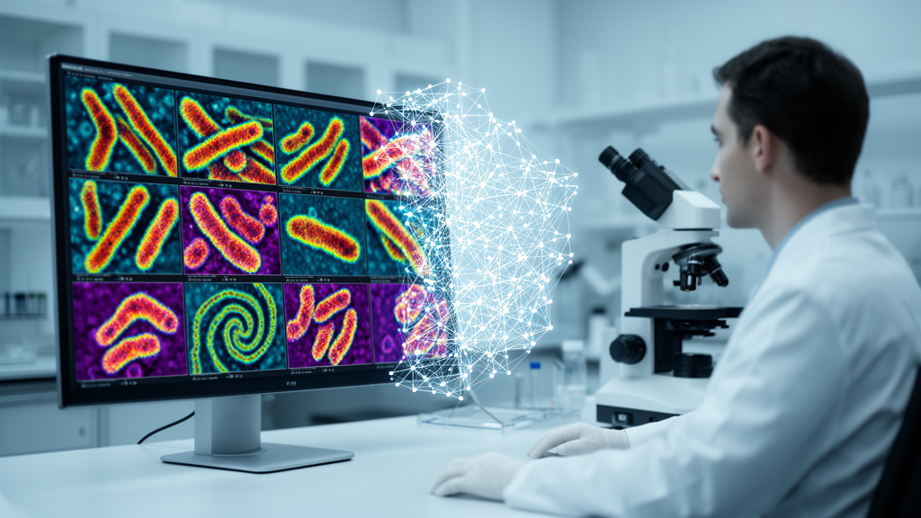
Identifying the exact species of bacterium lurking in a patient’s sample has long been a painstaking, human-dominated craft. Technicians peer through microscopes at Gram-stained slides, judging cell shapes, sizes, and staining patterns against years of accumulated experience. The process is subjective, slow, and vulnerable to fatigue, yet it remains a cornerstone of clinical microbiology. Now, a pair of researchers at Sambalpur University in Odisha, India, has unveiled a compact artificial intelligence system that can distinguish among 33 different bacterial species from ordinary microscopic images with accuracy exceeding 96 percent — and it does so fast enough, and small enough, to run on the kinds of inexpensive devices found in clinics far from any major laboratory.
The new framework, described in the journal Neural Computing and Applications, was built by Nitya Ranjan Manihira and Prabira Kumar Sethy of the university’s Department of Electronics. Their central challenge was one that plagues much of medical artificial intelligence: the available datasets of labeled bacterial microscopy images are small, and small data is notoriously unforgiving to deep neural networks, which typically demand enormous collections of examples before they learn to generalize. Rather than trying to grow a network from scratch, the team turned to transfer learning, a strategy in which a model pre-trained on millions of everyday images is repurposed for a specialized task, carrying with it a rich visual vocabulary already learned from its original training.
The backbone of their system is EfficientNetB0, a convolutional neural network architecture celebrated for squeezing remarkable accuracy out of very few parameters. On its own, however, a generic backbone can be easily distracted. Microscopy slides are messy places, littered with staining artifacts, debris, and uneven illumination that can masquerade as biological structure. To sharpen the model’s focus, the researchers bolted on a convolutional attention module — a mechanism inspired by the CBAM architecture that teaches the network, in effect, where to look. The attention module learns to weight both the spatial locations and the feature channels of the image, amplifying signals from regions dense with bacterial cells while suppressing the visual noise of the background.
The numbers behind the system tell a story of deliberate efficiency. The entire model contains just 3,761,747 trainable parameters, translating to an effective weight footprint of 14.35 megabytes — small enough to fit comfortably on a smartphone or embedded processor. Average inference time clocks in at 0.1457 seconds per image, equivalent to roughly 6.86 frames per second. In practical terms, that means the model can classify bacterial images nearly in real time on modest hardware, a property the authors highlight as making it suitable for edge devices and point-of-care settings where cloud computing and expensive workstations are unavailable.
Rigorous validation was central to the study’s design. The researchers employed five-fold stratified cross-validation, a technique that divides the data into five partitions and repeatedly trains and tests the model so that every image serves in both roles, while stratification preserves the class balance within each fold. On top of that, they held out a separate test set that the model never saw during training or tuning. The best-performing fold achieved a cross-validation accuracy of 95.44 percent with a standard deviation of 2.49 percent, and the model reached 96.43 percent accuracy on the untouched test set — a combination that suggests the network is genuinely generalizing rather than memorizing its training examples.
Class imbalance, one of the stubborn realities of biological datasets, received its own treatment. Some bacterial species in the dataset are represented by many images, while rarer species appear only sparingly, and naive models tend to ignore the underdogs entirely. The team countered this with class-balanced loss weighting, a training strategy that penalizes mistakes on rare classes more heavily, forcing the network to devote capacity to species it might otherwise overlook. The payoff is visible in the final metrics: the model achieved a macro F1-score of 0.964 and a weighted F1-score of 0.965 across all 33 taxa, with a perfect F1-score of 1.0 on 26 of the classes and still-reliable performance, at or above 0.667, on the under-represented species.
Perhaps the most clinically significant feature of the work is not the accuracy itself but the transparency that accompanies it. Deep learning models in medicine have long been criticized as black boxes, offering verdicts without reasons — a dealbreaker in a field where clinicians must justify decisions. To open the box, the researchers applied Grad-CAM, a visualization technique that uses the gradients flowing through the network to produce heatmaps showing which parts of the image drove each prediction. When overlaid on the original micrographs, these maps revealed that the attention mechanism was locking onto dense bacterial clusters and the discriminative morphological features that distinguish one species from another, while actively suppressing background artifacts. Each classification, in other words, comes with a spatially grounded explanation a microbiologist can inspect and judge.
The study builds on a publicly available dataset of bacteria images compiled for machine vision and digital biology research, and it situates itself within a rapidly growing body of work applying deep learning to microbiology. Earlier efforts have tackled bacterial colony classification, genus-level identification from hyperspectral microscopy, detection of drug-resistant cells in electron micrographs, and morphological diagnosis of bacterial vaginosis. What distinguishes the new work is the combination of scale — 33 classes rather than a handful — with an unusually light computational footprint and an explicit commitment to interpretability, three qualities that rarely coexist in a single system.
The implications for healthcare delivery could be substantial. In many parts of the world, trained microbiologists are scarce, and the delay between sample collection and species identification can stretch the time a patient spends on the wrong antibiotic. Gram staining has guided initial antibiotic therapy in clinical trials, underscoring how much hinges on rapid, accurate reading of stained slides. A lightweight model that runs at the point of care, explains its own reasoning, and performs reliably even on rare species could serve as a decision-support tool that augments, rather than replaces, human expertise — flagging likely species in seconds and letting technicians concentrate on the ambiguous cases.
Cautious optimism remains the appropriate posture. The model was trained and validated on a specific dataset of Gram-stained images, and real-world clinical deployment would demand testing across staining protocols, microscope models, and patient populations that vary far more widely than any single collection can capture. Still, the study demonstrates that the barriers of small data and limited compute — the two obstacles that most often keep laboratory AI confined to papers — can be overcome with careful architecture, disciplined validation, and a design philosophy that treats explanation as a feature rather than an afterthought. As attention-guided networks continue to shrink in size while growing in capability, the microscope slide may soon have a tireless, transparent second reader standing by.
Subject of Research: Attention-guided deep transfer learning for classifying 33 bacterial species from Gram-stained microscopic images
Article Title: Attention-guided deep transfer learning for 33-class bacterial species classification from microscopic images
Article References: Manihira, N. R., & Sethy, P. K. (2026). Attention-guided deep transfer learning for 33-class bacterial species classification from microscopic images. Neural Computing and Applications, 38(19), Article 779. https://doi.org/10.1007/s00521-026-12538-6
Image Credits: AI Generated
DOI: 10.1007/s00521-026-12538-6
Keywords: deep learning, transfer learning, bacterial classification, Gram stain, microscopy, attention mechanism, EfficientNetB0, Grad-CAM, explainable AI, clinical microbiology, point-of-care diagnostics, convolutional neural networks
News Source: Blake Davidson. (October 9, 2026). AI Learns to Spot 33 Bacterial Species From Microscope Images in a Fraction of a Second. Scienmag.