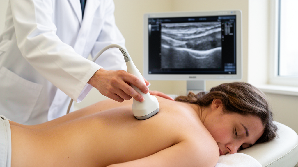
For decades, the effectiveness of a therapeutic massage has been judged largely by feel. A clinician presses, pulls, and glides their hands across a patient’s skin, and the verdict on whether the tissue has loosened rests on tactile intuition and the patient’s own description of their pain. That subjectivity has long frustrated researchers trying to understand what soft tissue manipulation actually does to the body’s mechanical landscape. Now, a team at the University of Virginia and Indiana University has shown that a trio of overhead cameras, a temporary skin tattoo, and a computer-vision technique borrowed from engineering can turn that intuition into hard numbers, revealing measurable changes in tissue mobility that align strikingly well with where patients report their pain.
The study, published in the Annals of Biomedical Engineering, focused on the cervicothoracic region, the junction between the neck and upper back where myofascial pain, a condition marked by tender trigger points and restricted movement, is especially common. Soft tissue manipulation, a family of massage-based interventions that includes myofascial release and instrument-assisted techniques, is widely used to treat this pain, yet no objective gold standard exists for assessing its effects. Existing tools such as myotonometry and shear-wave elastography measure stiffness only in small, localized spots and are sensitive to probe alignment, and they entirely miss the lateral gliding of fascial layers over one another, a property that many researchers believe is central to both restriction and relief.
The researchers’ solution was to watch the skin itself. They applied a semi-permanent speckle pattern to each participant’s upper back using custom tattoo stickers that react with skin proteins over 24 hours, producing a high-contrast pattern of dark blue dots roughly 1.5 millimeters across. The pattern works on all skin tones, darkening further on darker pigmentation, and remains visible for up to ten days before fading naturally. Three cameras mounted on a rotating arm above the massage table then recorded the speckled skin at 30 frames per second while an expert clinician performed a standardized pull test, dragging the tissue at a 45-degree angle to the myofascial plane in superior and inferior directions on both sides of the spine.
The imaging technique at the heart of the method is three-dimensional digital image correlation, or 3D-DIC, an optical approach that divides each frame into small pixel subsets and tracks their movement across stereo camera views to reconstruct full fields of displacement and strain. Using open-source software, the team computed eleven strain-based biomarkers from the resulting data, split into two families: tissue glide measures, which capture how far the skin and underlying tissue move in response to the clinician’s pull, and tissue deformation measures, which quantify how much the surface stretches or compresses under that load. Restricted tissue, the reasoning goes, behaves as if tethered, producing interrupted glide and elevated strain, while freed tissue should glide farther and deform less.
Nineteen generally healthy adults, ranging from 20 to 67 years old and reporting a spectrum of cervicothoracic pain, underwent the full protocol. Each received a 15-minute intervention delivered by a single expert clinician, combining five minutes of manual massage, including effleurage, cross-fiber strokes, petrissage, and skin rolling, with eight minutes of instrument-assisted soft tissue manipulation using a sensor-equipped device that recorded an average peak force of about 12 newtons and a stroke frequency near 1.5 hertz. Stretch assessments were repeated immediately before and after the intervention, with two additional participants assessed twice without any treatment to serve as a preliminary no-change control.
The aggregate results were unambiguous. Four biomarkers changed systematically after the intervention: maximum pull increased by 3.4 millimeters, from roughly 29.7 to 33.1 millimeters; maximum far-region displacement rose by 2.7 millimeters; ramp-on pull velocity climbed from about 33 to 42 millimeters per second; and total gross deformation increased as well. The velocity finding is particularly telling, because the clinician modulates each pull through tactile feedback toward a consistent target force. A faster ramp-up to that same end-feel implies the tissue offered less initial resistance, consistent with reduced viscoelastic stiffness after treatment. Pull propagation, the ratio of far to near displacement, held steady at around 65 percent, suggesting the extra motion was distributed proportionally across the tracked region.
At the individual level, the picture became even more compelling. Sixteen of the seventeen treated participants, 94 percent, showed measurable mobility changes after the intervention, and fifteen, or 88 percent, improved on at least one side of the body. Among the ten participants who reported a meaningful left-right pain asymmetry at baseline, meaning at least a two-point difference on a 0-to-10 scale, nine showed greater mobility gains on their more painful side. One representative participant, for instance, could have their tissue pulled 33 percent farther on the treated side after the intervention, at a 47 percent faster rate. The two untreated control participants showed no changes above the study’s conservative 25 percent threshold, and the classification held when the threshold was shifted to 20 or 30 percent.
Perhaps the most striking result concerned baseline asymmetries. Before any treatment, ten participants, just over half the cohort, exhibited bilateral mobility asymmetries detectable by the biomarkers. Eight of those ten also reported asymmetric pain, and in every single one of those eight cases, the less mobile side was the more painful side. This alignment echoes prior ultrasound findings that people with chronic low back pain show altered connective tissue thickness and reduced fascial shear strain, and it supports mechanistic accounts in which fascial densification, collagen cross-linking, reduced hydration, and diminished hyaluronan-mediated lubrication between layers increase stiffness and nociceptive drive together.
The authors are careful about what the biomarkers actually measure. The optical signals capture a composite response of skin, subcutaneous tissue, fascial layers, and deeper muscle, not fascial gliding in isolation, and the modest 3.4-millimeter gain in maximum pull should be read in context: sub-millimeter changes in cervical facet joint gapping during spinal manipulation have previously been linked to meaningful improvements in range of motion and pain. The study also has limits. It involved a single clinician, a small cohort, and no sham control, and mobility was assessed only immediately before and after treatment, so the durability of the gains remains unknown. One participant whose mobility declined after treatment despite reporting no pain asymmetry highlights that psychological stress can raise localized muscle tone, a reminder that mechanical and symptomatic change do not always move in lockstep.
Even so, the implications for clinical practice are considerable. Two biomarkers, total gross deformation and ramp-on pull velocity, proved the most sensitive across participants, suggesting a compact toolkit could preserve diagnostic power while simplifying implementation. The semipermanent speckle pattern, visible for ten days, opens the door to tracking how a single treatment evolves over days or how multiple sessions accumulate. The team’s next steps include a sham-controlled design, a larger and more diverse cohort, and a blinded two-clinician setup to separate treatment effects from assessment bias. If those efforts succeed, the humble massage could finally acquire something it has always lacked: a ruler.
Subject of Research: Optical measurement of skin surface strain to quantify changes in soft tissue mobility after massage-based treatment of myofascial pain
Article Title: Detecting Intervention-Specific Change in Soft Tissue Mobility Aligned with Regional Pain Through Optically Measured Skin Surface Strains
Article References: Kao, A. R., Loghmani, M. T., & Gerling, G. J. (2026). Detecting Intervention-Specific Change in Soft Tissue Mobility Aligned with Regional Pain Through Optically Measured Skin Surface Strains. Annals of Biomedical Engineering. https://doi.org/10.1007/s10439-026-04373-6
Image Credits: AI Generated
DOI: 10.1007/s10439-026-04373-6
Keywords: digital image correlation, soft tissue manipulation, myofascial pain, fascia, skin strain, biomarkers, manual therapy, tissue mobility, biomechanics, massage, cervicothoracic region, rehabilitation
News Source: Ophelia Keating. (October 9, 2026). Cameras Catch Massage Loosening Stiff Tissue on the More Painful Side of the Body. Scienmag.