One of the most puzzling patterns in Alzheimer’s disease research is that inheritance from a mother appears to matter more than inheritance from a father. People whose mothers had dementia face a substantially elevated risk compared with those whose fathers were affected, and they tend to show earlier onset, faster cognitive decline, and heavier burdens of amyloid-beta protein in the brain, even before any memory problems appear. Yet family history is surprisingly easy to lose: parents may die young, diagnoses may never have been made, and relatives’ medical records may be incomplete. A new study published in PLOS Aging Health by Olivia Veatch of Children’s Mercy Research Institute and Robyn Honea of the University of Kansas Medical Center, together with their colleagues, tackles this gap with a machine learning approach that attempts to reconstruct a person’s maternal family history from brain scans and mitochondrial DNA alone.
The research team drew on two of the largest repositories in American dementia research: the National Alzheimer’s Coordinating Center, which aggregates standardized clinical evaluations from 40 federally funded Alzheimer’s Disease Research Centers, and NIAGADS, the National Institute on Aging’s genetics storage service. From these databases they assembled a cohort of 1,129 participants with known family history, with a mean age of 69 years, 61 percent of whom were women. Forty-four percent reported a mother or maternal relative with dementia, mild cognitive impairment, or Alzheimer’s disease, while the remainder reported either only paternal history or no family history at all, with both parents living past age 60 and cognitively normal. About 71 percent of participants had normal cognition at the time of imaging, and roughly 12 percent met criteria for dementia.
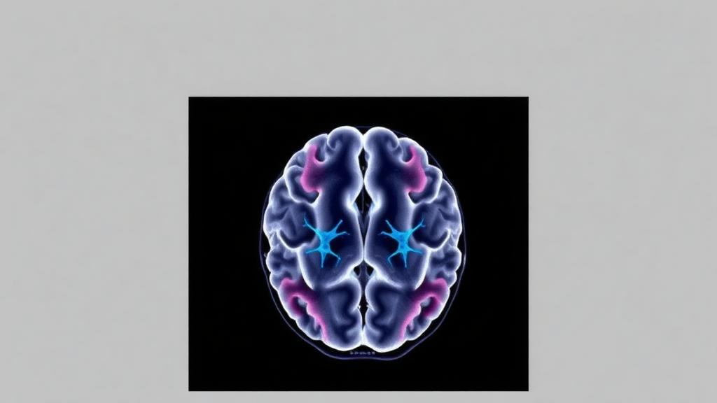
The predictive features came from two biologically motivated sources. The first was structural magnetic resonance imaging, processed with the automated segmentation tool FreeSurfer, yielding nine carefully chosen measures: the hippocampal occupancy score, which reflects shrinkage of the hippocampus relative to the inferior lateral ventricle; white matter hyperintensity volume, a marker of cerebrovascular damage; and normalized volumes of seven cortical and subcortical regions, including the precuneus, entorhinal cortex, lingual gyrus, inferior and superior temporal gyri, inferior parietal cortex, and fusiform gyrus. The second source was mitochondrial DNA, the small circular genome inherited almost exclusively through the maternal line. Using HaploGrep 2 software, the researchers classified each participant’s mitochondrial haplogroup, an inherited branch on the maternal phylogenetic tree that reflects ancient ancestry and can influence mitochondrial function.
The machine learning pipeline was rigorous. The data were split 80:20 into training and testing sets using stratified sampling to preserve the proportion of family-history-positive cases, and hyperparameters were tuned with grid search combined with four-fold cross-validation. Seven algorithms competed: decision tree, logistic regression, naive Bayes, linear and radial support vector machines, linear discriminant analysis, and XGBoost, a gradient-boosted ensemble method that builds many decision trees sequentially and corrects each one’s errors. Models were compared on accuracy and the area under the receiver operating characteristic curve, and feature importance was assessed through a rank-based integration of two complementary metrics: how frequently XGBoost used each feature across its tree ensemble, and the discriminative range of each feature’s conditional probabilities in the naive Bayes model.
The results were modest but statistically meaningful. Among imaging-only models, XGBoost performed best, reaching an accuracy and AUC of 0.59, with sensitivity of about 0.56 and specificity of 0.62; naive Bayes followed closely at 0.58. A label-shuffling permutation test, in which outcome labels were randomly permuted 1,000 times to generate a null distribution, confirmed that the XGBoost model performed significantly better than chance, with an empirical p-value of 0.006. The standout feature was the volume of the precuneus, a large region on the inner surface of the parietal lobes, followed by the hippocampal occupancy score, entorhinal cortex, lingual gyrus, and temporal regions. The least informative features were the inferior parietal cortex, fusiform gyrus, and white matter hyperintensity volume.
The precuneus is a compelling winner. It serves as a central hub of the brain’s default mode network, the constellation of regions most active during rest and introspection, and it is among the earliest and most heavily affected areas in Alzheimer’s disease. Previous work by the same group and others has shown that cognitively normal people with a maternal family history display altered gray matter volume, progressive atrophy in the precuneus and medial temporal cortex over two years, abnormal default mode network activity centered on this region, and elevated amyloid-beta deposition specifically in the precuneus and posterior parietal and cingulate cortices. Postmortem studies add a mechanistic thread: reduced activity of the choline acetyltransferase enzyme in the precuneus has been linked to increased amyloid binding, higher soluble amyloid-beta 42 levels, and more advanced Alzheimer’s pathology.
The mitochondrial story is more nuanced. When the researchers added haplogroup classifications to the imaging data, the sample shrank by roughly 36 percent to 717 participants, because not everyone had genetic data available. Smaller samples usually degrade predictive performance, yet the combined model held its ground: XGBoost achieved an accuracy and AUC of 0.62, with sensitivity of 0.61 and specificity of 0.63. The R0 intermediate clade emerged as the most influential mitochondrial feature, though the precuneus remained the single most important variable overall. The authors are candid that the added value of the mitochondrial data remains unclear, since feature importance indicated minimal contribution relative to imaging. Still, the fact that performance did not collapse despite the much smaller sample suggests the haplogroups carry some signal worth pursuing.
The mechanistic rationale for looking at mitochondria at all is rooted in maternal inheritance. Because mitochondrial DNA passes almost exclusively from mother to child, it offers a natural explanation for why maternal, but not paternal, family history elevates amyloid accumulation independently of APOE epsilon 4 status, the strongest common genetic risk factor. Earlier studies found reduced cytochrome oxidase activity in adult children of mothers with Alzheimer’s disease but not fathers, and postmortem analyses have linked lower mitochondrial genome copy numbers in the brain to increased odds of Alzheimer’s neuropathology. Notably, the predictive models deliberately excluded APOE epsilon 4 status to avoid problems with incorporating ordinal variables, and when the team statistically adjusted for age, sex, race, ethnicity, and APOE genotype before modeling, performance dropped for most algorithms, indicating that some of the discriminative signal reflects demographic and genetic differences between the family-history groups rather than a purely imaging-based signature.
The authors are appropriately cautious about clinical translation. Repeated analyses across ten random seeds yielded mean AUCs near 0.53, revealing limited stability, and restricting the sample to self-reported white participants reduced performance further. The cohort was overwhelmingly of European ancestry, limiting generalizability to populations with different haplogroup distributions, and family history relied on self-report, which is vulnerable to recall bias. The cross-sectional design cannot establish whether precuneus differences precede or follow other disease changes. The researchers emphasize that these findings should not be used for individual risk prediction, and they flag the ethical concerns of communicating probabilistic susceptibility without definitive interventions. What the study does deliver is a framework: proof of concept that multimodal data can partially recover maternal family history when it is unknown, a reinforcement of the precuneus as the signature region of maternally inherited risk, and a roadmap toward larger, longitudinal, ancestrally diverse studies that incorporate rare mitochondrial variants and copy number. If that promise is fulfilled, the invisible half of a family tree may one day be read from a scan and a blood sample.
Subject of Research: Machine learning prediction of maternal family history of Alzheimer's disease using structural neuroimaging and mitochondrial haplogroups
Article Title: Using machine learning of neuroimaging and mitochondrial haplogroups to predict maternal family history of Alzheimer’s disease
Article References: Veatch, O. J., Dutta, S., Mansel, C. O., Townley, R., Sardiu, M. E., & Honea, R. A. (2026). Using machine learning of neuroimaging and mitochondrial haplogroups to predict maternal family history of Alzheimer’s disease. PLOS Aging and Health, 1(3), e0000045. https://doi.org/10.1371/journal.page.0000045
Image Credits: AI Generated
DOI: 10.1371/journal.page.0000045
Keywords: Alzheimer's disease, maternal family history, machine learning, XGBoost, precuneus, mitochondrial DNA, haplogroups, structural MRI, neuroimaging, APOE epsilon 4, default mode network, risk prediction
News Source: Cassandra Pierce. (October 8, 2026). Brain Scans and Mitochondrial DNA Offer Clues to Maternal Alzheimer’s Risk. Scienmag.