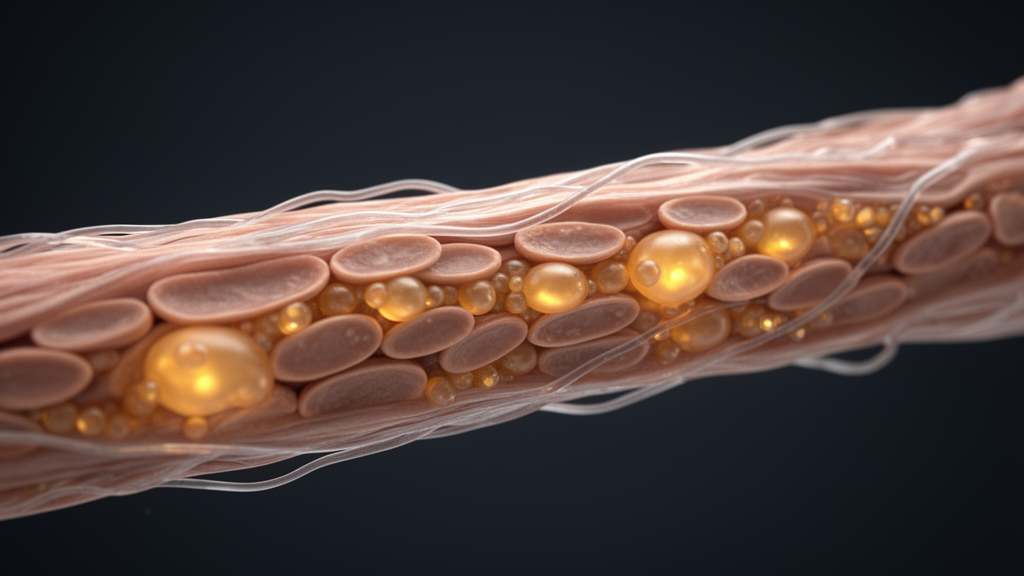
Deep inside every muscle fiber, embedded in the folds of the mitochondria that power movement, sits a peculiar fat molecule that scientists have long known is essential for life but rarely blamed for the visible toll of growing old. A new study published in Nature Aging argues that this lipid, called cardiolipin, may be one of the hidden puppet masters of muscle aging. When its levels fall, as they do in both aging mice and aging humans, skeletal muscle undergoes a striking transformation: fast, powerful glycolytic fibers shrink and give way to smaller, slower, oxygen-hungry oxidative fibers. The researchers show that this is not a passive consequence of wear and tear but an active, genetically orchestrated program, driven by a signaling cascade that runs from the mitochondrial membrane all the way to the cell’s nucleus.
The paradox that motivated the work is one that has puzzled muscle biologists for decades. Aging is well known to sap mitochondrial function, reducing the capacity of cells to generate energy through respiration. Yet at the same time, aging muscle becomes enriched in the very fiber types that are most dependent on mitochondria. Fast-twitch fibers, which rely on rapid glycolytic metabolism for bursts of force, selectively atrophy, while slow-twitch oxidative fibers persist. Something must be coordinating this shift, and whether failing mitochondria are merely a bystander or an active instigator has remained unresolved. The new research, led by Fabian Finger and Zachary Gerhart-Hines at the University of Copenhagen together with an international team, provides the strongest evidence yet that a single mitochondrial lipid sits near the top of that causal chain.
Cardiolipin is no ordinary fat. It is found almost exclusively in the inner mitochondrial membrane, where its four fatty acyl chains and distinctive shape confer the tight curvature that cristae, the energy-generating folds of the membrane, require. It stabilizes the respiratory supercomplexes that shuttle electrons through the electron transport chain, supports the enzymes of oxidative phosphorylation, and participates in virtually every aspect of mitochondrial architecture and function. Because cardiolipin is so central, its disruption has been implicated in conditions ranging from cardiomyopathy to neurodegeneration and Barth syndrome, a rare genetic disorder. All cardiolipin species originate from a single rate-limiting enzymatic reaction catalyzed by cardiolipin synthase 1, encoded by the gene Crls1, which made the enzyme an obvious point of attack for the researchers.
The team began by asking whether cardiolipin declines with age. Comparing young mice at nine weeks with mice at two years, they measured cardiolipin levels in both the oxidative soleus muscle and the glycolytic extensor digitorum longus, and found that total cardiolipin fell similarly in both muscle types. The gene most significantly reduced in aged muscle was Crls1 itself. Crucially, the same pattern appeared in human muscle biopsies: total cardiolipin declined in older participants aged 53 to 69 compared with young adults aged 22 to 35, along with significant shifts in the composition of individual cardiolipin species and reduced expression of cardiolipin synthesis genes. The conservation across species suggested that the loss of this lipid is a genuine signature of muscle aging rather than a rodent quirk.
To test whether the decline is a cause rather than a consequence of aging, the researchers genetically deleted Crls1 specifically in the skeletal muscle of young mice, creating a model they call MUCKO. The deletion reproduced a global reduction in cardiolipin across muscle types that closely mirrored the pattern seen in aged animals, even though cardiolipin represents less than two percent of the muscle phospholipidome. The consequences were dramatic. MUCKO mice failed to gain lean mass after weaning, and although oxidative muscle is naturally more cardiolipin-rich, it was the glycolytic EDL muscle that suffered the most severe atrophy. Transmission electron microscopy revealed disrupted cristae architecture in both muscle types, and respiratory supercomplex formation was uniformly diminished. When respiration was corrected for mitochondrial content, NADH-linked respiration, oxidative phosphorylation capacity, succinate-linked respiration and complex IV activity were all significantly reduced, confirming that cardiolipin loss cripples mitochondrial function across fiber types.
Yet the most surprising finding was not the damage but the adaptation. Despite losing respiratory capacity, the glycolytic EDL muscle of MUCKO mice dramatically increased its mitochondrial content, with citrate synthase activity and COX IV staining rising to levels resembling the oxidative soleus. Transcriptomic and proteomic profiling showed that EDL was undergoing a global fast-to-slow fiber-type switch: the most glycolytic type IIb fibers decreased while more oxidative IIa and IIx subtypes expanded. Individual fiber cross-sectional areas shrank, but the total number of fibers was unchanged, indicating that atrophy of existing fibers, not cell death, drove the tissue’s decline. In short, deleting one lipid-synthesizing enzyme in young muscle recapitulated a constellation of aging hallmarks on an accelerated timescale, establishing cardiolipin deficiency as a plausible causal driver of the aging phenotype.
The hunt for the molecular messenger connecting membrane to nucleus led to estrogen-related receptor gamma, or ERRγ, a nuclear receptor already known for its role in exercise-induced oxidative remodeling. In MUCKO muscle, Esrrg expression rose specifically in glycolytic EDL, mirroring the pattern of mitochondrial mass, and it was also elevated in aged mouse muscle. In cultured muscle cells, depleting Crls1 selectively increased the oxidative myosin heavy chain isoforms I and IIa, but this induction was completely blocked either by knocking down ERRγ with interfering RNA or by treating cells with the pharmacological antagonist GSK5182. Genome-wide, of the 773 genes significantly regulated by Crls1 loss, 626 proved dependent on ERRγ activity. The receptor also bound the promoter of myogenin, a master regulator of muscle differentiation, placing ERRγ upstream of MYOG in a signaling hierarchy that ultimately rewrites the muscle’s contractile machinery. This is retrograde communication in its purest form: a change in the lipid composition of the mitochondrial inner membrane is transduced into a transcriptional program that reshapes the entire fiber.
Why would aging muscle deliberately remodel itself this way? The answer, the researchers found, lies in antioxidant defense. Cardiolipin-deficient muscle showed elevated mitochondrial reactive oxygen species production and increased lipid peroxidation, along with a near-universal upregulation of ROS defense proteins. Proteomic and metabolomic analyses revealed that glucose entering these muscles was being rerouted away from complete glycolysis and into the pentose phosphate pathway, the hexosamine biosynthetic route, serine metabolism and the folate cycle, all of which generate the reducing power needed to neutralize oxidative stress. Isotopic tracing confirmed increased conversion of glucose to serine, and the muscles consumed markedly more glucose in vivo, making the mice strikingly glucose tolerant despite their mitochondrial dysfunction. When the researchers gave mice the antioxidant N-acetylcysteine for eight weeks, glucose uptake in the cardiolipin-deficient muscles was blunted, suggesting that ROS itself stimulates the sugar hunger that fuels these protective pathways.
Perhaps the most provocative result concerns reversibility. Using an inducible knockout model in adult mice, the team showed that stopping the genetic ablation allowed modest re-expression of Crls1, which raised cardiolipin levels from 27 percent to 67 percent of normal. That partial recovery was enough to initiate reversal of the dramatic weight loss and muscle atrophy, and it completely rescued the premature mortality that otherwise killed the knockout mice, apparently from diaphragm dysfunction. Even more counterintuitively, chronic antioxidant supplementation made things worse, lowering the sarcopenic index and further activating the stress response, presumably by starving the muscle of the glucose flux its defense pathways require. The findings reframe the fiber-type shift of aging not as degeneration alone but as an adaptive, ERRγ-driven survival strategy, one that cannot be patched by simply swallowing antioxidants. If future work confirms that boosting cardiolipin synthesis in aged muscle can restore youthful fiber composition, the humble mitochondrial fat may become an unexpected target in the quest for healthier aging.
Subject of Research: Role of the mitochondrial membrane lipid cardiolipin in driving age-related skeletal muscle fiber-type adaptations via estrogen-related receptor γ signaling
Article Title: Mitochondrial membrane lipid cardiolipin controls fiber-type adaptations in aging muscle via estrogen-related receptor γ
Article References: Finger, F., Frost, M., Watanabe, S., Ma, T., Kang, T., Biswas, D., Zapata-Perez, R., Anton, V., Dollet, L., Markussen, L. K., Senoo, N., Karavaeva, I., Basse, A. L., Dmytriyeva, O., TeSlaa, T., Havelund, J. F., Ehrlich, A. M., Moreno-Torres, M., Yonamine, C. Y., … Gerhart-Hines, Z. (2026). Mitochondrial membrane lipid cardiolipin controls fiber-type adaptations in aging muscle via estrogen-related receptor γ. Nature Aging. https://doi.org/10.1038/s43587-026-01227-7
Image Credits: AI Generated
DOI: 10.1038/s43587-026-01227-7
Keywords: cardiolipin, mitochondria, skeletal muscle, aging, sarcopenia, ERRγ, Crls1, fiber-type switching, mitonuclear signaling, reactive oxygen species, glucose metabolism, Nature Aging
News Source: Beatrice Stafford. (October 8, 2026). A Fading Mitochondrial Fat May Drive the Way Aging Muscles Change Shape. Scienmag.