Gastric cancer remains one of the world’s most lethal malignancies, and one of its most dangerous features is something surgeons cannot see with their own eyes: lymphovascular invasion, or LVI, the moment tumor cells slip into lymphatic vessels and blood vessels and gain a highway to spread elsewhere in the body. Once LVI is present, the risk of recurrence climbs and long-term outcomes worsen. Yet in most clinical practice today, doctors only learn whether a patient’s tumor has invaded these vessels after surgery, when a pathologist dissects the removed tissue under a microscope. By then, the treatment decisions that could have been shaped by that knowledge have already been made.
A new study published in BMC Cancer by Jian Shang, Yaolu Li, Lidan Yang, Donghui Zhang and colleagues at the Affiliated Cancer Hospital of Guangzhou Medical University set out to change that timeline. The research team developed and internally validated a machine-learning model that combines standard clinical variables with quantitative features extracted from multiparametric magnetic resonance imaging to predict LVI before a patient ever enters the operating room. The work, a retrospective single-center study, also explored whether the model’s output could stratify patients by their risk of disease-free survival, offering a glimpse of how preoperative imaging intelligence might one day guide gastric cancer management.
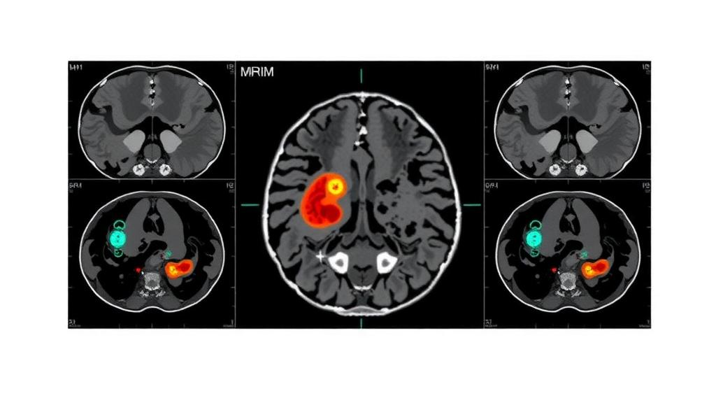
The technical foundation of the study is radiomics, a field that converts the pixel-and-voxel patterns inside medical images into hundreds or thousands of numerical descriptors. Where a radiologist’s eye might notice that a tumor looks heterogeneous or ill-defined, a radiomics pipeline quantifies exactly how heterogeneous, how textured, how intense, and how variable the tumor’s signal is across different imaging sequences. The researchers drew on three complementary MRI sequences: fat-suppressed T2-weighted imaging, which highlights tissue water content and edema; apparent diffusion coefficient maps derived from diffusion-weighted imaging, which reflect the restricted movement of water molecules in densely packed tumor tissue; and venous-phase contrast-enhanced MRI, which reveals how the tumor takes up gadolinium-based contrast agent through its blood supply.
Each sequence captures a different facet of tumor biology. Restricted diffusion, for example, often corresponds to high cellularity, a hallmark of aggressive malignancies. Contrast enhancement patterns can reflect the chaotic, leaky neovasculature that tumors build to feed themselves. T2 signal characteristics can distinguish mucin-rich or necrotic regions from solid cellular components. By mining all three sequences simultaneously, the team aimed to build a digital fingerprint of the tumor that correlates with the microscopic reality of vascular invasion.
The study population comprised 458 patients with gastric cancer, randomly assigned to a training cohort of 320 patients and an internal test cohort of 138. This split is a critical methodological safeguard: models built and evaluated on the same patients routinely overstate their own accuracy, so holding out a test set provides a more honest estimate of performance. From the MRI data, the researchers extracted radiomics features and applied feature reduction techniques, including least absolute shrinkage and selection operator regression, known as LASSO, which penalizes complexity and drives the coefficients of uninformative features to zero. After this filtering process, six radiomics features and four clinical predictors survived as the final model inputs.
The team then constructed three logistic-regression models to compare different predictor sets, designating the combined clinical-radiomics model as the primary model. Logistic regression may seem modest in an era of deep neural networks, but it offers transparency, calibration, and robustness with modest sample sizes, qualities that matter enormously in clinical prediction. Secondary comparisons using alternative machine-learning algorithms, including support vector machines and linear discriminant analysis, were performed with fold-restricted random oversampling, a technique that balances the classes within each cross-validation fold to avoid data leakage while addressing the inevitable imbalance between LVI-positive and LVI-negative patients.
The headline result: the combined clinical-radiomics model achieved an area under the receiver operating characteristic curve, or AUC, of 0.775 in the training cohort and 0.752 in the internal test cohort, with a bootstrap 95 percent confidence interval of 0.663 to 0.840. An AUC of 0.75 places the model in the territory of moderate discrimination, meaning it performs meaningfully better than chance but is far from infallible. At a sensitivity-oriented threshold of 0.217, chosen in the training set using the Youden index, the model detected LVI with a test sensitivity of 0.795, catching roughly four out of five invasion-positive cases, while its negative predictive value reached 0.855. In practical terms, when the model says a tumor is unlikely to have lymphovascular invasion, that reassurance is correct about 86 percent of the time.
Calibration, the question of whether predicted probabilities match observed reality, is often neglected in machine-learning studies but received careful attention here. In the test cohort, the calibration intercept was 0.092, the slope 0.965, and the Brier score 0.180, all indicating that the model’s probability estimates were reasonably honest rather than systematically overconfident or underconfident. Decision-curve analysis further assessed the clinical utility of the model across a range of threshold probabilities, evaluating the net benefit a patient population would gain if decisions were made according to the model’s predictions.
Perhaps the most intriguing finding is exploratory: the model’s output appeared to carry prognostic weight. Using an X-tile-derived cutoff of 0.59 established in the training data and applied unchanged to the test cohort, the researchers divided patients into risk groups for disease-free survival. In an adjusted complete-case Cox proportional hazards model, each standard deviation increase in the model’s predicted probability was associated with a hazard ratio of 1.57 for worse disease-free survival, with a 95 percent confidence interval of 1.21 to 2.03. A single preoperative number, computed from scans and blood markers, appeared to encode information about how the disease would unfold, independent of the adjustment variables included in the analysis.
The authors are appropriately measured about what their model can and cannot do. They emphasize that the model is not ready for stand-alone clinical use and requires prospective external validation in independent centers before any deployment. The study was retrospective and single-center, meaning the MRI scanners, imaging protocols, and patient demographics of one institution shaped the data. Radiomics features are notoriously sensitive to variations in acquisition parameters, and a model trained in Guangzhou may not transfer cleanly to a hospital with different equipment. The disease-free survival findings, too, are explicitly labeled exploratory, and the sensitivity analyses using a training-median cutoff underscore the uncertainty around how the risk stratification should be operationalized.
Still, the study adds to a growing body of evidence that the information needed to characterize a tumor’s aggressiveness may already be hiding inside routine imaging. Every gastric cancer patient undergoing staging typically receives cross-sectional imaging; the marginal cost of running a radiomics algorithm on those existing scans is nearly zero. If future prospective studies confirm these results, a high negative predictive value at a sensitivity-oriented threshold suggests a plausible triage role: patients whose scans argue strongly against LVI might be candidates for less aggressive perioperative planning, while those flagged as high risk could be prioritized for intensified neoadjuvant therapy, extended lymph node dissection, or closer surveillance. The vision is not to replace pathologists but to move critical biological knowledge upstream in the treatment timeline, from the postoperative pathology report to the preoperative clinic visit.
The work also highlights a broader trend in oncology: the convergence of interpretable machine learning, multiparametric imaging, and clinical data into decision-support tools that are auditable rather than opaque. By retaining logistic regression as the primary model and reporting calibration alongside discrimination, the Guangzhou team modeled the kind of methodological discipline that regulatory science increasingly demands. The path from a 0.75 AUC in a single center to a validated clinical tool is long, but this study maps an early stretch of it, showing that the vessels a tumor invades in secret may leave traces visible to an algorithm long before any scalpel is lifted.
Subject of Research: Preoperative prediction of lymphovascular invasion in gastric cancer using multiparametric MRI radiomics and machine learning
Article Title: Preoperative prediction of lymphovascular invasion and prognostic risk stratification in gastric cancer using multiparametric MRI radiomics and machine learning: a retrospective model development and internal validation study
Article References: Preoperative prediction of lymphovascular invasion and prognostic risk stratification in gastric cancer using multiparametric MRI radiomics and machine learning: a retrospective model development and internal validation study. (n.d.). https://doi.org/10.1186/s12885-026-17042-7
Image Credits: AI Generated
DOI: 10.1186/s12885-026-17042-7
Keywords: gastric cancer, lymphovascular invasion, multiparametric MRI, radiomics, machine learning, logistic regression, disease-free survival, predictive modeling, medical imaging, cancer prognosis, internal validation, LASSO
News Source: Nathaniel Bowman. (October 7, 2026). AI Reads MRI Scans to Predict Hidden Gastric Cancer Spread Before Surgery. Scienmag.