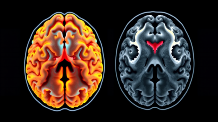
Going without food for days does more than shrink the waistline. It quietly reshapes the way the brain reacts to the emotional world around it, and it does so in stages that follow a strikingly predictable timetable. That is the central message of a new longitudinal study published in the Journal of Translational Medicine, in which a team of Chinese researchers tracked the electrical activity of nineteen healthy adults as they completed a fifteen-day, medically supervised total fast. Using high-density electroencephalography, the researchers captured millisecond-by-millisecond snapshots of how the participants’ brains evaluated disturbing images before the fast, twice during it, and again after two days of recovery eating. The result is one of the most detailed timelines yet of how sustained metabolic deprivation alters human emotional reactivity, and it suggests that the brain does not simply become more or less reactive as energy reserves drain away. Instead, it appears to pass through distinct adaptive phases, each with its own neural signature.
The study, led by Qian Wu and Wenbin Fu of the Second Affiliated Hospital of Guangzhou University of Chinese Medicine, together with Jiajin Yuan of Sichuan Normal University and colleagues, was designed around a deceptively simple question: what happens to the brain’s rapid emotional alarm system when the body is forced to run on nothing but water? Prolonged fasting exposes a person to a sustained energy deficit and a chronic psychological stress load, both of which are known in the broader literature to influence how people process negative and threatening information. Yet most previous work has relied on single snapshots, comparing fasted individuals with non-fasted controls at one moment in time. By measuring the same participants repeatedly across four phases, the team could watch the adaptation unfold within each brain rather than inferring it from differences between people.
The measurement tool at the heart of the study is the event-related potential, or ERP, a technique that averages the brain’s electrical responses to repeated stimuli recorded from the scalp. Because ERPs unfold over tens to hundreds of milliseconds, they reveal stages of processing that unfold far too quickly for conscious report. The researchers used a negative emotional picture Oddball task, a classic paradigm in which participants view a stream of images and occasionally encounter an unexpected, emotionally charged deviant. The deviant automatically pulls attention, and the brain’s response to it can be decomposed into well-characterized components. The occipital N1, arising roughly 100 milliseconds after stimulus onset over visual cortex, reflects early attentional resource recruitment. The fronto-central N2, peaking around 200 to 300 milliseconds, indexes attentional orienting to unexpected or conflicting information. The parietal P3, the latest of the three, is widely read as a marker of motivational salience and evaluative processing, the brain’s judgment of how much an event matters.
Alongside the EEG recordings, the team collected supplementary measures of autonomic flexibility. Before each Oddball session, participants rested while the researchers recorded photoplethysmograms, an optical measure of blood volume pulse, and respiration traces. From these they computed respiratory sinus arrhythmia, or RSA, the natural speeding and slowing of the heart that is synchronized with the breathing cycle. RSA is commonly interpreted as an index of parasympathetic vagal influence and, in the emotional domain, of the flexibility with which a person’s physiology can respond to changing emotional contexts. Lower RSA during a challenging period is generally taken to reflect reduced capacity for adaptive emotional engagement. Including this autonomic layer gave the researchers a second, independent line of evidence against which to check the patterns emerging from the scalp recordings.
Nineteen healthy adults carried the study through its demanding schedule. Each completed the emotional Oddball task at four points: at baseline before fasting began, on day seven of complete fasting, on day fourteen of complete fasting, and on day two of the recovery period, when normal eating had resumed. The entire experimental process was conducted under full medical supervision, with a professional medical team monitoring participants’ physical and mental condition throughout, and the protocol was approved by a biomedical research ethics committee in accordance with the Declaration of Helsinki. All participants provided informed consent. The longitudinal design allowed the researchers to analyze the data with linear mixed-effect models, a statistical framework that respects the repeated-measures structure of the data and estimates how each phase differs from baseline within the same individuals.
The ERP results revealed a phase-dependent pattern that the authors describe as a shift in the architecture of emotional reactivity. Early in the fast, at day seven, the dominant change was an amplification of the parietal P3, the component associated with salience evaluation. In other words, one week into total food deprivation, the participants’ brains appeared to assign heightened motivational significance to the negative emotional deviants, treating the disturbing images as more consequential than they had at baseline. This heightened evaluative response is consistent with the intuition that an energized alarm system accompanies the early stages of a metabolic challenge, when the body’s stress physiology is mobilized and the brain may be primed to prioritize threat-relevant information.
By day fourteen, however, the picture had changed. The later phase of the fast was characterized not by amplified evaluation but by an attenuated fronto-central N2, the component reflecting attentional orienting to unexpected information. After two weeks without food, the participants’ brains appeared less responsive to the surprise value of the emotional deviants, suggesting a blunting of the orienting response rather than a continued amplification of it. Meanwhile, across both fasting phases, at day seven and day fourteen, the participants showed increased occipital N1 responses to the explicit task demands, indicating greater recruitment of early attentional resources whenever the task required it. The brain, in this reading, was still capable of mobilizing visual attention when instructed, but its automatic, involuntary reactions to emotional surprise had shifted from amplification to dampening as the fast wore on.
Crucially, the story did not end with the fast. On day two of recovery, after participants had resumed eating, all of the observed ERP changes had returned to baseline levels. The heightened salience evaluation of the early phase and the attenuated orienting of the late phase were both reversible, disappearing once normal energy balance was restored. The autonomic data told a complementary story: RSA estimates supported a reduction in emotional context flexibility during the complete fasting period, again consistent with the idea that sustained metabolic stress constrains the physiological flexibility that supports adaptive emotional responding. The convergence of a cortical measure and a peripheral autonomic measure on the same conclusion strengthens the overall pattern, even though the two signals were collected in different states, one during active task performance and one at rest.
The authors are careful to frame the work as hypothesis-generating rather than definitive. With nineteen participants and no mention of a control group consuming a normal diet across the same schedule, the findings require replication in larger controlled studies before the identified neural and autonomic patterns can be considered for practical use. The researchers themselves emphasize that the ERP and RSA readouts should not yet be treated as validated monitoring tools. Still, the study points toward an intriguing application. If the phase of a fast can be read out from a person’s neural and autonomic responses, then neurobehavioral monitoring strategies could in principle be timed to the metabolic state of the individual, deploying countermeasures, whether nutritional, psychological, or pharmacological, at the moments when the brain’s emotional machinery is known to shift gear. Such considerations matter in contexts ranging from clinical fasting protocols to extreme occupational settings, though the authors stop short of claiming readiness for any of these uses.
What makes the study compelling beyond its immediate numbers is the conceptual shift it forces. Emotional reactivity under starvation is often assumed to move in a single direction, toward either irritability and hypervigilance or apathy and blunting. This work suggests the reality is a choreographed sequence: an early phase in which the brain magnifies the significance of negative events, followed by a later phase in which automatic orienting to the unexpected fades, all while the capacity for deliberate attentional effort is preserved and all of it unwinding within days of refeeding. The brain, it seems, does not merely degrade under metabolic stress; it adapts in stages, trading one configuration of emotional processing for another as the energy crisis deepens, and then quietly reverting when the crisis passes. Disentangling the mechanisms behind that choreography, from ketone metabolism to stress hormones to sleep disruption, is the task the authors now hand to the field.
Subject of Research: Neural and autonomic adaptation of emotional reactivity during prolonged complete fasting, measured longitudinally with event-related potentials
Article Title: Phase-dependent adaptation of emotional reactivity during prolonged complete fasting: longitudinal ERP evidence from a human metabolic challenge
Article References: Wu, Q., Fan, Z., Lin, D., Wang, S., Sun, L., Liang, X., Wen, X., Wu, B., Lin, W., Wang, T., Yuan, J., & Fu, W. (2026). Phase-dependent adaptation of emotional reactivity during prolonged complete fasting: longitudinal ERP evidence from a human metabolic challenge. Journal of Translational Medicine. https://doi.org/10.1186/s12967-026-09011-2
Image Credits: AI Generated
DOI: 10.1186/s12967-026-09011-2
Keywords: complete fasting, emotional reactivity, event-related potentials, P3, N2, N1, respiratory sinus arrhythmia, EEG, metabolic stress, neurobehavioral monitoring, longitudinal study, Journal of Translational Medicine
Cite Scienmag News
APA
MLA
Chicago
Cassandra Pierce. (September 25, 2026). Fasting Rewires the Brain’s Emotional Radar in Two Distinct Phases, EEG Study Finds. Scienmag. https://scienmag.com/fasting-rewires-the-brains-emotional-radar-in-two-distinct-phases-eeg-study-finds/
Cassandra Pierce. “Fasting Rewires the Brain’s Emotional Radar in Two Distinct Phases, EEG Study Finds.” Scienmag, 25 September 2026, https://scienmag.com/fasting-rewires-the-brains-emotional-radar-in-two-distinct-phases-eeg-study-finds/. Accessed 25 September 2026.
Cassandra Pierce. “Fasting Rewires the Brain’s Emotional Radar in Two Distinct Phases, EEG Study Finds.” Scienmag. September 25, 2026. https://scienmag.com/fasting-rewires-the-brains-emotional-radar-in-two-distinct-phases-eeg-study-finds/
Copy citation
Download RIS
Tags: brain reactivity phases during food deprivationcomplete fastingEEGEEG-based neural response to fastingemotional reactivityevent-related potentialsFasting effects on brain emotional processingfasting-induced neural plasticity and emotional regulationhigh-density EEG in fasting researchimpact of total fasting on emotional radarJournal of Translational Medicinelongitudinal EEG study on fasting and brain functionlongitudinal studymetabolic deprivation and emotional reactivitymetabolic stressN1N2neural mechanisms of fasting and emotion regulationneural signatures of fasting-induced emotional changesneurobehavioral monitoringP3respiratory sinus arrhythmiastages of brain adaptation during prolonged fastingtemporal dynamics of brain response to fasting