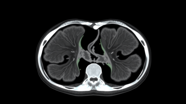
Medical image segmentation, the task of teaching computers to trace the exact outlines of organs and lesions inside CT scans, MRIs, and endoscopic images, sits at the heart of modern computer-aided diagnosis. Radiation oncologists use these digital contours to plan treatments, surgeons rely on them to navigate around critical structures, and clinical trials depend on them to measure whether a tumor is shrinking. Yet the deep learning models that perform this work have long been caught in a trade-off. Convolutional neural networks, the workhorses of medical imaging for the past decade, are efficient and good at detecting local textures and edges, but they struggle to capture long-range relationships, the kind of whole-organ context needed to understand where one structure ends and another begins. Transformer-based architectures solve that context problem with self-attention, but their computational cost grows quadratically with the number of image patches, making them expensive and sometimes impractical for the high-resolution volumes used in hospitals.
A research team at Shaanxi University of Technology in Hanzhong, China, led by corresponding author Chuanghua Yang, has now introduced a new architecture designed to escape this dilemma. In a study published in the journal Complex & Intelligent Systems, the researchers present REF-VMUNetUp, a network built on the Mamba architecture, a recently popularized state-space model that processes sequences with linear rather than quadratic complexity. The work builds on VM-UNet, a well-known pure-Mamba segmentation network, but the authors argue that VM-UNet leaves important performance on the table. Their new model targets three specific weaknesses: inadequate modeling of positional information, degradation of translation invariance during upsampling, and inaccurate segmentation of organ boundaries.
To understand why position matters, it helps to consider what Mamba actually does. Originally developed for natural language processing, Mamba treats an image as a sequence of tokens and sweeps through them with a selective state-space mechanism, deciding dynamically which information to remember or forget as it goes. This gives it the long-range modeling power of a Transformer at a fraction of the computational cost, which is precisely why it has generated such excitement in the medical imaging community. The catch is that, in its basic form, the mechanism is largely indifferent to where each token sits in the image. Two patches with similar appearance can be treated nearly identically even if one belongs to the liver and the other to the right kidney, and that ambiguity can blur the very boundaries clinicians care about most.
REF-VMUNetUp addresses this with a two-dimensional rotary position embedding, which the authors call 2D-RoPE. Rotary position embeddings first gained fame in large language models, where they encode the relative distance between tokens by rotating feature vectors in the complex plane; the angle of rotation depends on position, so the attention-like scoring between two tokens naturally reflects how far apart they are. The Shaanxi team adapted this idea to two dimensions, encoding both row and column coordinates so that the network can represent relative spatial relationships across the image plane. The result, according to the study, is a marked improvement in the model’s ability to represent relative position information, which in turn sharpens its grasp of anatomical layout without adding the heavy cost of explicit positional attention.
The second innovation tackles a subtler problem: upsampling. Segmentation networks follow an encoder-decoder design, compressing the image into abstract feature maps and then expanding them back to full resolution to produce pixel-level labels. The expansion step, or upsampling, is traditionally done with fixed interpolation rules such as bilinear or nearest-neighbor resampling, which treat every location in the image the same way. That uniformity erodes translation invariance and produces edge blurring, because a smooth interpolation cannot know that a sharp organ boundary happens to pass through a particular neighborhood. The researchers’ answer is a module they call D-UP, short for dynamic upsampling, which predicts offsets dynamically for each sampling location and reorganizes channels accordingly. In effect, the network learns where to place each output pixel, warping the resampling grid to follow contours rather than ignoring them, which mitigates the edge blurring that fixed upsampling introduces.
The third component operates on the skip connections, the pathways that carry fine-grained detail from the encoder directly to the decoder. In classic U-Net-style architectures, these connections are what allow the final segmentation to retain precise edges, but they also pass along low-level noise and redundant features. REF-VMUNetUp inserts a module named L-HFP, a lightweight high-frequency processing unit, into the skip pathway. Its job is to adaptively enhance high-frequency features, the rapidly varying components of the signal that correspond to edges, fine textures, and thin structures. By amplifying exactly the information the decoder needs to capture critical details such as organ boundaries and tumor margins, and suppressing what it does not, the module improves the fidelity of the final output without a large computational overhead.
The team evaluated the architecture on the Synapse multi-organ dataset, a widely used benchmark of abdominal CT scans in which algorithms must delineate organs including the aorta, gallbladder, kidneys, liver, pancreas, and spleen. Performance was measured with two standard metrics: the Dice similarity coefficient, or DSC, which quantifies the overlap between the predicted segmentation and the ground truth, and HD95, the 95th-percentile Hausdorff distance, which captures how far the worst boundary errors stray from the true contour. REF-VMUNetUp improved mean DSC by 1.83 percentage points and reduced HD95 by 2.09 millimeters relative to the baseline comparison, gains that are meaningful in a field where fractions of a millimeter can influence treatment planning. The reduction in HD95 is particularly telling, because that metric is dominated by the largest boundary errors, precisely the failures the dynamic upsampling and high-frequency modules were designed to fix.
Impressive results on a single benchmark can reflect overfitting, so the researchers also tested generalization on three additional public datasets spanning different imaging modalities and clinical tasks: ACDC, which features cardiac MRI for measuring heart function; ISIC2018, a dermoscopy dataset for skin lesion segmentation; and CVC-ClinicDB, containing colonoscopy images of polyps. The model’s consistent performance across these varied domains suggests that the benefits of position-aware Mamba scanning and dynamic upsampling are not tied to one anatomy or scanner type. The authors note that all datasets used in the research are publicly available and that they declare no conflict of interest, and the work was supported by the National Natural Science Foundation of China, the Key Research and Development Program of Shaanxi Province, and a Qinchuangyuan Scientists and Engineers team grant.
The broader significance of the study lies in what it says about the direction of medical AI. For years, the field has been pulled between the efficiency of convolutions and the context awareness of Transformers, and Mamba-style state-space models promise a way to have both. But as this work demonstrates, simply swapping a Transformer for a Mamba block is not enough; the architecture must also respect the geometric and structural priors of imaging, from relative position to sharp boundaries, or it will forfeit much of its potential. By embedding rotary position encoding, learned dynamic resampling, and adaptive high-frequency enhancement into a linear-complexity backbone, REF-VMUNetUp offers a template for how that can be done. If such models continue to mature, the practical payoff could be faster, cheaper, and more precise automated contouring, bringing reliable multi-organ and lesion segmentation closer to routine clinical use in hospitals that cannot afford heavyweight Transformer infrastructure.
Subject of Research: A Mamba-based deep learning architecture for medical image segmentation
Article Title: REF-VMUNetUp: a position-aware mamba network with dynamic upsampling for medical image segmentation
Article References: Yang, C., Yang, H., Yuan, H., Gao, W., Li, W., Pan, Z., Li, Y., & Bai, X. (2026). REF-VMUNetUp: a position-aware mamba network with dynamic upsampling for medical image segmentation. Complex & Intelligent Systems. https://doi.org/10.1007/s40747-026-02515-w
Image Credits: AI Generated
DOI: 10.1007/s40747-026-02515-w
Keywords: medical image segmentation, Mamba, state space models, VM-UNet, rotary position embedding, dynamic upsampling, deep learning, computer vision, multi-organ segmentation, Synapse dataset, U-Net, computational efficiency
Cite Scienmag News
APA
MLA
Chicago
Blake Davidson. (September 24, 2026). Mamba-Based AI Sharpens Medical Image Segmentation With Position Awareness and Dynamic Upsampling. Scienmag. https://scienmag.com/mamba-based-ai-sharpens-medical-image-segmentation-with-position-awareness-and-dynamic-upsampling/
Blake Davidson. “Mamba-Based AI Sharpens Medical Image Segmentation With Position Awareness and Dynamic Upsampling.” Scienmag, 24 September 2026, https://scienmag.com/mamba-based-ai-sharpens-medical-image-segmentation-with-position-awareness-and-dynamic-upsampling/. Accessed 24 September 2026.
Blake Davidson. “Mamba-Based AI Sharpens Medical Image Segmentation With Position Awareness and Dynamic Upsampling.” Scienmag. September 24, 2026. https://scienmag.com/mamba-based-ai-sharpens-medical-image-segmentation-with-position-awareness-and-dynamic-upsampling/
Copy citation
Download RIS
Tags: clinical applications of medical image segmentationcomputational efficiencycomputational efficiency in medical image analysiscomputer visionconvolutional neural networksdeep learningdeep learning in medical imagingdynamic upsamplingdynamic upsampling in medical image analysishigh-resolution medical image processinglong-range context capture in medical imagesMambaMamba-based AI architecturemedical image segmentationmulti-organ segmentationposition awareness in segmentation modelsREF-VMUNetUp neural networkrotary position embeddingstate-space modelsSynapse datasettransformer-based architectures for medical imagingU-NetVM-UNet