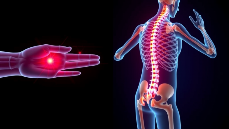
One of the most disabling and poorly understood consequences of spinal cord injury may finally have a molecular explanation. Researchers report that adrenomedullin, a small peptide hormone released following injury to the spinal cord, acts as the critical messenger that drives heterotopic ossification, the abnormal formation of mature bone inside skeletal muscle and other soft tissues. The study, published in Experimental & Molecular Medicine, maps a previously unrecognized neuro–immune–bone axis that connects the damaged central nervous system to inflammatory cells and, ultimately, to bone-forming progenitors trapped in muscle. The findings suggest that a single circulating signal, arriving from the injured cord itself, can reprogram the local tissue environment so thoroughly that muscle begins to behave like a bone-forming organ.
Heterotopic ossification affects a substantial fraction of people with spinal cord injury, with estimates in clinical literature ranging from around 20 percent to more than 60 percent depending on how aggressively patients are screened. The condition typically emerges below the level of the lesion, most often around the hips, knees and elbows, and produces swelling, stiffness, progressive joint immobility and, in severe cases, complete ankylosis of the affected joint. For individuals already coping with paralysis, the added loss of joint range can devastate independence, interfering with seating, transfers, dressing and the ability to use a wheelchair comfortably. Current treatment options are limited and unsatisfying: nonsteroidal anti-inflammatory drugs and bisphosphonates are used prophylactically with mixed evidence, radiation therapy is occasionally deployed in high-risk cases, and surgical excision remains the definitive intervention, despite high recurrence rates and considerable operative risk.
What has been missing is a coherent account of how a wound in the spinal cord communicates with skeletal muscle distant from the injury site. Inflammation at the muscle level has long been implicated, and macrophages, the versatile immune cells that flood damaged tissue, have been suspected participants. Bone morphogenetic protein signaling, particularly through BMP2 and related ligands, is known to be a powerful inducer of ectopic bone. Yet none of these observations explained the striking temporal and anatomical link to the central nervous system injury itself. The new work addresses that gap by identifying adrenomedullin as the long-range signal, and by showing that it does not act alone but through a choreographed interaction between the nervous system, the immune system and mesenchymal progenitors residing in muscle.
Adrenomedullin is a 52-amino-acid peptide first isolated from human pheochromocytoma tissue in the early 1990s and subsequently recognized as a near-universal stress messenger. It is produced by vascular smooth muscle, endothelial cells, epithelia and neurons, and it participates in vasodilation, angiogenesis, immune modulation and tissue repair. Its receptors, composed of the calcitonin receptor-like receptor paired with receptor activity-modifying proteins, are widely distributed. In the context of spinal cord injury, the researchers found that adrenomedullin expression rises markedly in the injured cord, and that the peptide reaches the circulation and downstream tissues at levels capable of instructing cells far from the lesion. This elevation is not a passive byproduct of tissue damage; the study demonstrates that it is functionally required for the ectopic bone program to begin.
The mechanistic core of the paper lies in what adrenomedullin does once it arrives in muscle. Using rodent models of spinal cord injury combined with pharmacological blockade and genetic approaches, the team showed that adrenomedullin recruits and polarizes macrophages toward a pro-inflammatory, pro-osteogenic state. These macrophages, in turn, become a rich local source of bone morphogenetic protein 2, the master osteoinductive cue. Meanwhile, adrenomedullin acts directly on mesenchymal stromal cells and muscle-resident progenitors, sensitizing them to the BMP2 signal and pushing them along an osteoblastic differentiation pathway. The result is a feed-forward circuit: the neural signal summons and shapes the immune response, the immune response supplies the osteogenic ligand, and the sensitized progenitors execute the bone-forming program. The authors describe this as a neuro–immune–bone axis, a three-node signaling architecture in which each node is necessary for the pathology to unfold.
The experimental evidence supporting this model is notably multi-layered. In mice subjected to spinal cord injury, heterotopic bone formed in paralyzed limbs on a timeline consistent with the human clinical course, and the extent of ossification correlated with circulating adrenomedullin. Neutralizing the peptide with antibodies, antagonizing its receptors, or depleting macrophages each substantially reduced ectopic bone formation. Conversely, administering exogenous adrenomedullin to animals with otherwise insufficient neural drive promoted macrophage accumulation and osteogenic differentiation in muscle. Conditional genetic strategies further clarified the cellular division of labor: macrophage-derived BMP2 proved essential, while the osteogenic competence of the mesenchymal compartment depended on adrenomedullin receptor signaling. Transcriptional profiling of the affected muscle revealed enrichment of inflammatory, angiogenic and osteogenic gene programs, consistent with a tissue being actively converted from a muscle identity toward a bone-forming niche.
Perhaps the most clinically resonant finding concerns timing and reversibility. The adrenomedullin-driven cascade was shown to be active during a defined window after cord injury, and interventions that blocked the axis during that window prevented or markedly attenuated heterotopic ossification. This reframes the condition not as an inevitable consequence of paralysis but as a targetable signaling disease, one that could in principle be intercepted in the days to weeks following injury, before the first radiographic sign of bone appears. Because adrenomedullin antagonists and anti-BMP2 strategies already exist in various experimental and clinical forms, the pathway offers a plausible translational route. The caveat, which the authors acknowledge, is that adrenomedullin performs beneficial functions in vascular integrity, wound healing and immune regulation, so systemic blockade carries risks. Local delivery strategies, short treatment windows, or agents tuned to the specific receptor subtype involved may be needed to separate therapeutic benefit from collateral harm.
The study also carries broader implications beyond spinal cord medicine. Heterotopic ossification arises in other settings, including traumatic brain injury, severe burns, blast injuries in military personnel, and genetic conditions such as fibrodysplasia ossificans progressiva. In several of these contexts, central nervous system trauma or systemic inflammatory storms are part of the clinical picture, raising the possibility that neuro–immune–bone communication is a general principle of ectopic bone formation rather than a peculiarity of cord injury. If adrenomedullin or related neural signals contribute to ossification after burns or brain injury, the therapeutic horizon widens considerably. Even in fibrodysplasia ossificans progressiva, where an activating ACVR1 mutation provides the primary osteogenic driver, inflammatory flares are known to precipitate bone episodes, and understanding how neural and immune signals amplify that driver could inform combination therapies.
From a basic science perspective, the work adds to a growing recognition that the nervous system is not merely a bystander in regenerative and pathological processes but an active endocrine organ whose injury products reshape distant tissues. The concept of a neuro–immune–bone axis echoes earlier discoveries of neuroimmune interactions in pain, cachexia and bone homeostasis, but here the axis is shown to operate in a pathologically constructive direction, building ectopic skeleton rather than maintaining existing bone. It also underscores the macrophage’s dual identity: the same cells that clear debris and support repair can be commandeered by a circulating peptide into serving as local factories for osteoinductive ligands. Dissecting which macrophage subsets respond to adrenomedullin, and whether their polarization can be selectively redirected, represents an obvious next step.
For the millions of people worldwide living with spinal cord injury, and for the clinicians who manage the secondary complications of paralysis, the identification of a single, druggable molecular trigger for heterotopic ossification is a genuine advance. It converts a decades-old clinical observation, that paralyzed limbs sometimes grow bone, into a defined signaling pathway with named nodes, testable intermediates and established pharmacological levers. Clinical translation will require careful validation in human tissue and trials that respect the peptide’s physiological roles, but the conceptual shift is already complete: heterotopic ossification after spinal cord injury is now understood as a systemic endocrine event, initiated in the damaged cord, amplified by the immune system, and executed by progenitors in muscle that never asked to become bone cells.
Subject of Research: Adrenomedullin-driven heterotopic ossification after spinal cord injury via a neuro–immune–bone axis
Article Title: Adrenomedullin from spinal cord injury drives heterotopic ossification in skeletal muscle via a neuro–immune–bone axis
Article References: Chen, J., Wang, Z., Zeng, X., Wu, X., Li, G., Miao, N., Deng, Y., Zhang, D., Chen, X., Lai, H., Wan, Y., Wang, L., & Li, X. (2026). Adrenomedullin from spinal cord injury drives heterotopic ossification in skeletal muscle via a neuro–immune–bone axis. Experimental & Molecular Medicine. https://doi.org/10.1038/s12276-026-01836-7
Image Credits: AI Generated
DOI: 10.1038/s12276-026-01836-7
Keywords: spinal cord injury, heterotopic ossification, adrenomedullin, macrophages, BMP2, neuro–immune–bone axis, mesenchymal progenitors, ectopic bone, osteogenesis, Experimental & Molecular Medicine, spinal, cord
Cite Scienmag News
APA
MLA
Chicago
Cassandra Pierce. (September 23, 2026). Spinal Cord Injury Signal Found to Trigger Bone Growth in Muscle. Scienmag. https://scienmag.com/spinal-cord-injury-signal-found-to-trigger-bone-growth-in-muscle/
Cassandra Pierce. “Spinal Cord Injury Signal Found to Trigger Bone Growth in Muscle.” Scienmag, 23 September 2026, https://scienmag.com/spinal-cord-injury-signal-found-to-trigger-bone-growth-in-muscle/. Accessed 23 September 2026.
Cassandra Pierce. “Spinal Cord Injury Signal Found to Trigger Bone Growth in Muscle.” Scienmag. September 23, 2026. https://scienmag.com/spinal-cord-injury-signal-found-to-trigger-bone-growth-in-muscle/
Copy citation
Download RIS
Tags: adrenomedullinadrenomedullin role in bone formationBMP2circulating signals in injury responsecordectopic boneExperimental & Molecular Medicineheterotopic ossificationheterotopic ossification pathophysiologyinflammation and bone developmentinjury-induced bone growthmacrophagesmesenchymal progenitorsmuscle-to-bone transformation processneuro-immune-bone signaling axisneuro–immune–bone axisneuroimmune interactions in skeletal abnormalitiesosteogenesissoft tissue calcification in paralysissoft tissue ossification after spinal injuryspinalSpinal Cord Injuryspinal cord injury molecular mechanismstherapeutic targets for heterotopic ossification