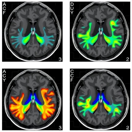
A quiet revolution is unfolding in how physicians understand one of the most consequential blood vessel blockages in the human brain. New research published in BMC Medical Imaging reveals that two tiny territories deep within the brain, fed by branches no wider than a human hair, respond in strikingly different ways to a blocked middle cerebral artery—and recover differently once that blockage is reopened. The finding, based on detailed CT perfusion imaging in patients undergoing endovascular recanalization, offers clinicians a fresh lens on the hidden hemodynamics of chronic stroke-causing lesions and may help refine which patients stand to benefit most from reopening procedures.
The study, led by Yujie Sun and colleagues at the Shengli Clinical Medical College of Fujian Medical University and the Affiliated Hospital of Qingdao University, focused on patients with symptomatic non-acute middle cerebral artery occlusion, or MCAO—a condition in which the major artery supplying blood to large portions of the brain has been blocked for more than two weeks, yet patients continue to experience symptoms. Unlike the frantic timelines of acute stroke care, where clot-busting drugs and thrombectomy dominate, non-acute occlusions present a more ambiguous clinical landscape. The brain has had time to adapt, collateral vessels may have sprouted, and the question of whether restoring flow through the blocked artery will actually help becomes far less straightforward.
At the heart of the research lies a delicate vascular architecture known as the lenticulostriate arteries, or LSAs. These are small, thin perforating vessels that branch directly off the M1 segment of the middle cerebral artery and plunge deep into the brain to nourish critical structures, including the internal capsule and portions of the basal ganglia—regions responsible for movement, sensation, and the relay of motor signals. The LSAs are traditionally divided into two groups: the medial lenticulostriate arteries, which arise more proximally and tend to be larger, and the lateral lenticulostriate arteries, which arise slightly further along the vessel and fan out laterally. Because these vessels are end arteries—meaning they have virtually no backup supply from neighboring vessels—damage or hypoperfusion in their territories can cause devastating, often permanent deficits.
What makes the new study compelling is its territory-specific approach. Rather than treating the deep brain as a uniform block of tissue, the researchers used CT perfusion imaging to separately quantify blood flow dynamics in the medial and lateral LSA territories, comparing the affected side of the brain with the healthy contralateral side. CT perfusion works by rapidly injecting iodinated contrast while the CT scanner repeatedly images the same brain region, tracking how the contrast washes in and out of tissue. From these dynamics, four key parameters are derived: cerebral blood volume (CBV), the amount of blood present in a given volume of tissue; cerebral blood flow (CBF), the rate at which blood moves through the tissue; mean transit time (MTT), the average time blood takes to pass through the capillary bed; and time to peak (TTP), how long it takes contrast concentration to reach its maximum.
To account for natural side-to-side variation, the team calculated relative indices—rCBV, rCBF, rMTT, and rTTP—by comparing each territory on the affected side with its mirror image on the healthy side. This normalization strategy is crucial in perfusion imaging, because absolute values can vary between individuals and between scanners, while relative asymmetries often reveal pathology more reliably. Eighteen patients who underwent successful endovascular recanalization, confirmed by the modified Thrombolysis in Cerebral Infarction (mTICI) grading on digital subtraction angiography, and who had perioperative CT perfusion scans, were retrospectively analyzed with paired statistical comparisons pre-specified before the analysis.
The pre-treatment findings painted a picture of two territories living very different realities under the same arterial occlusion. The lateral LSA territory showed the more severe insult: its cerebral blood volume and cerebral blood flow were significantly reduced compared with the healthy side, while its mean transit time and time to peak were markedly prolonged. In plain terms, less blood was arriving, and what did arrive moved sluggishly through the tissue. The medial territory, by contrast, told a subtler story. Although its blood flow was reduced and its transit times delayed—clear signs of compromised supply—its cerebral blood volume remained preserved. Blood was arriving slowly, but the tissue was still holding a near-normal quantity of blood within its vessels.
This dissociation between preserved volume and reduced flow is physiologically meaningful, and it points to compensatory vasodilation. When perfusion pressure drops, the cerebral vasculature has a first line of defense: the small arterioles dilate to reduce resistance and maintain flow. As pressure falls further, flow declines, but the dilated vessels still hold blood, keeping CBV relatively stable even as CBF falls and transit times lengthen. In this framework, the medial territory appeared to be sitting in a state of exhausted-but-active compensation—its reserve mobilized, its vessels maximally widened, its blood volume defended even as delivery faltered. The lateral territory, having lost ground on all fronts, suggested a deeper stage of hemodynamic compromise, where even the compensatory mechanisms were no longer sufficient to defend tissue perfusion.
Why would the lateral territory fare worse than the medial one? The authors note that the two groups of perforators may possess different hemodynamic vulnerabilities. The medial lenticulostriate arteries arise more proximally on the M1 segment, potentially closer to collateral inflow and at a different angle relative to the direction of occlusive flow, while the lateral branches, arising slightly downstream and oriented differently, may be more sensitive to pressure drops across the occluded segment. Anatomical differences in vessel caliber, branching pattern, and the territory each group supplies may compound these pressures. Whatever the precise anatomical basis, the imaging data demonstrate that deep perforator territories cannot be treated as a single hemodynamic unit in chronic MCA occlusion.
Then came the intervention: endovascular recanalization, in which physicians reopen the blocked artery—typically using a combination of catheter navigation, stent placement, and balloon angioplasty—to restore antegrade flow through the previously occluded vessel. After successful reopening, the follow-up CT perfusion scans documented meaningful changes in both territories, but with distinct signatures. The lateral LSA territory showed the more classical pattern of successful reperfusion: relative cerebral blood flow increased, and relative mean transit time and time to peak shortened, indicating that blood was once again arriving briskly. This is the profile clinicians hope to see when they reopen an occluded artery—faster flow, faster transit, restored supply.
The medial territory also showed improved flow and shortened transit times, confirming that recanalization benefited it as well. But one parameter moved in the opposite direction: relative cerebral blood volume decreased after treatment. Rather than indicating deterioration, this decrease is consistent with the resolution of compensatory vasodilation. Once the occlusion is lifted and perfusion pressure is restored, the chronically dilated arterioles no longer need to remain maximally open; vascular tone returns toward normal, vessel caliber narrows, and blood volume within the tissue falls accordingly. In this reading, the drop in rCBV is not a warning sign but a physiological sigh of relief—a measurable marker that the brain’s emergency adaptations are being stood down.
The clinical implications of these differential patterns are potentially significant. First, they suggest that territory-based CT perfusion analysis could become a more precise tool for assessing hemodynamic vulnerability before deciding on intervention in non-acute MCAO. Patients whose lateral LSA territories show severe hypoperfusion with exhausted reserve may represent those most likely to benefit from recanalization, while the state of the medial territory’s compensatory response might inform expectations about recovery. Second, the post-treatment profiles offer a template for evaluating whether a procedure has genuinely improved deep perforator hemodynamics—something conventional angiography, which shows the parent vessel, cannot directly assess. Recanalizing the artery is one thing; confirming that blood flow dynamics in the vulnerable deep territories have actually normalized is another, and CT perfusion provides that window.
The study also carries a broader conceptual message: the deep brain is not monolithic. Perforator territories that sit side by side, supplied by branches of the same parent artery, can occupy different positions along the spectrum of hemodynamic compensation. This territory-specific framing may prove relevant not only to chronic MCA occlusion but also to other conditions involving perforator vessels, including small-vessel disease and the notorious risk of periprocedural complications during endovascular interventions, where reperfusion injury or hemorrhage in perforator territories remains a feared event.
The researchers are appropriately measured about the scope of their conclusions. With eighteen patients analyzed retrospectively, the findings are best viewed as hypothesis-generating, laying the groundwork for larger prospective studies that could validate territory-specific perfusion metrics as predictors of outcome. The authors also emphasize that their paired, pre-specified comparison design strengthens the internal validity of the observed changes, since each patient served as their own anatomical control before and after treatment. The work was funded by grants from the Fujian Province Joint Funds for the Innovation of Science and Technology, and the study was conducted under the approval of the Ethics Committee of the Affiliated Hospital of Qingdao University in accordance with the Declaration of Helsinki.
For now, the message for the field is clear. When a major brain artery has been quietly blocked for weeks, the tiny territories it once fed are not merely surviving or dying as one. The medial lenticulostriate territory may be straining under compensatory vasodilation, its vessels dilated to their limits, while the lateral territory slips toward more profound hypoperfusion. And when flow is restored, the brain’s recovery writes itself in perfusion numbers—flow rising, transit quickening, and blood volume gently falling as the machinery of compensation powers down. Territory-specific CT perfusion, the study suggests, can read both the struggle and the relief, opening a path toward more individualized decisions in the treatment of chronic cerebral arterial occlusion.
Subject of Research: Territory-specific CT perfusion changes in the medial and lateral lenticulostriate artery territories before and after endovascular recanalization in patients with symptomatic non-acute middle cerebral artery occlusion
Subject of Research: Medicine
Article Title: Differential patterns of territory-specific CT perfusion reflecting hemodynamic status before and after revascularization in non-acute MCA occlusion
Article References: Sun, Y., Ye, P., Wang, Z., Zhang, Y., & Li, Y. (2026). Differential patterns of territory-specific CT perfusion reflecting hemodynamic status before and after revascularization in non-acute MCA occlusion. BMC Medical Imaging. https://doi.org/10.1186/s12880-026-02732-2
Image Credits: AI Generated
DOI: 10.1186/s12880-026-02732-2
Keywords: Middle cerebral artery occlusion, Recanalization, CT perfusion, Lenticulostriate artery, Neuroimaging, Cerebral blood flow, Cerebral blood volume, Mean transit time, Endovascular treatment, Deep perforator territories
Cite Scienmag News
APA
MLA
Chicago
Cassandra Pierce. (September 3, 2026). Territory-specific CT perfusion tracks blood flow changes after chronic MCA revascularization. Scienmag. https://scienmag.com/territory-specific-ct-perfusion-tracks-blood-flow-changes-after-chronic-mca-revascularization/
Cassandra Pierce. “Territory-specific CT perfusion tracks blood flow changes after chronic MCA revascularization.” Scienmag, 3 September 2026, https://scienmag.com/territory-specific-ct-perfusion-tracks-blood-flow-changes-after-chronic-mca-revascularization/. Accessed 3 September 2026.
Cassandra Pierce. “Territory-specific CT perfusion tracks blood flow changes after chronic MCA revascularization.” Scienmag. September 3, 2026. https://scienmag.com/territory-specific-ct-perfusion-tracks-blood-flow-changes-after-chronic-mca-revascularization/
Copy citation
Download RIS
Tags: blood flow changesbrain blood flow changesbrain blood supply dynamicsbrain hemodynamicsbrain perfusion analysiscerebral hemodynamicsChronic MCA occlusionChronic MCA revascularizationcollateral circulation in strokecollateral vessel adaptationCT perfusion imagingdeep brain vascular territoriesendovascular recanalizationendovascular revascularizationischemic stroke imagingnon-acute middle cerebral artery occlusionnon-acute stroke treatmentreperfusion therapy outcomesstroke patient selectionstroke recoverystroke treatment strategiesterritory-specific blood flowterritory-specific stroke recovery