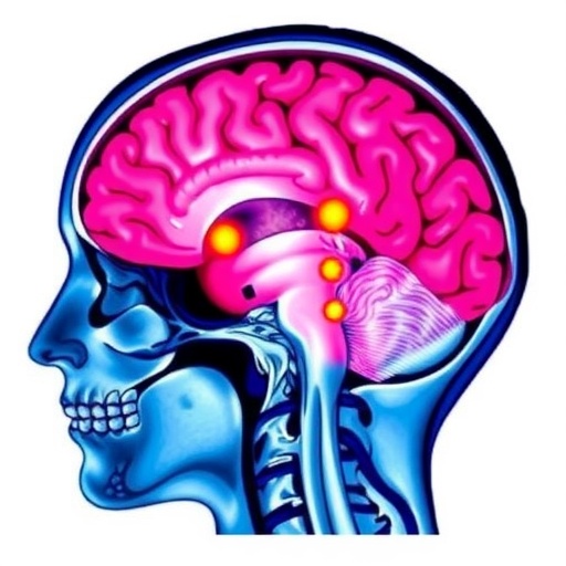
Her Mind Went Silent for Two Months — Then Doctors Found Antibodies Attacking Her Brain
For two months, a 60-year-old woman watched — in whatever way she still could — as her mind quietly slipped away. She had been healthy her entire life, with no psychiatric history, no neurological illness, and no obvious trigger. Yet her thinking slowed, her behavior turned strange, and the people around her could no longer reach her. By the time she arrived at the hospital, she had stopped speaking altogether, a condition known as mutism, and was making purposeless movements that served no visible goal. There was no stroke, no tumor, and no virus detectable anywhere in her body. The culprit, a new case report reveals, was her own immune system, which had turned against a molecule her brain cannot function without: the N-methyl-D-aspartate receptor, one of the central switches of learning, memory, and consciousness.
The case, published in the March 2026 issue of Annals of Clinical and Translational Neurology, describes what its authors diagnosed as cerebral cortical encephalitis in anti-NMDA receptor (anti-NMDAR) autoimmune encephalitis — a form of brain inflammation in which antibodies target the NMDA receptor, a glutamate-gated ion channel concentrated at the excitatory synapses of the cerebral cortex. When neurologists examined the woman, they found impaired consciousness and negativistic behavior, meaning she actively resisted instructions and interaction. More striking were stereotyped orolingual dyskinesias: repetitive, unvarying movements of the lips, jaw, and tongue, performed over and over without intention or control. These involuntary grimacing and chewing motions are a hallmark of anti-NMDAR encephalitis. They emerge when the disease disrupts the cortical and basal ganglia circuits that normally keep movement smooth, purposeful, and quiet — circuits that depend heavily on precisely the receptor under attack.
Brain imaging immediately complicated the picture. Magnetic resonance imaging with T2-fluid-attenuated inversion recovery (FLAIR) sequences — a technique that suppresses the bright signal of cerebrospinal fluid so that inflammation stands out against a dark background — revealed hyperintense lesions in the bilateral frontal–parietal regions and the left temporal lobe. Critically, the abnormalities traced the leptomeninges, the delicate arachnoid and pia mater membranes that envelope the brain and carry blood vessels along its surface. Diffusion-weighted imaging (DWI), which detects the restricted movement of water molecules within swollen cells, showed corresponding signal changes in the same territories. Leptomeningeal hyperintensity is classically associated with meningitis, metastatic cancer, or inflammatory disease of the membranes themselves, so its appearance in a suspected autoimmune encephalitis was genuinely unusual. Bilateral, multilobar leptomeningeal involvement of this kind is rarely reported in anti-NMDAR disease, making the scan simultaneously the most alarming and the most instructive clue in the entire work-up.
A lumbar puncture delivered the decisive evidence. The woman’s cerebrospinal fluid contained 28 white blood cells per microliter, 95 percent of them lymphocytes — a mild lymphocytic pleocytosis, the classic fingerprint of inflammation inside the central nervous system. Protein and glucose were both normal, a pattern that argues strongly against bacterial infection. Most important, testing for anti-NMDAR antibodies came back strongly positive, with a cerebrospinal fluid titer of 1:100 compared with a serum titer of only 1:10. That steep gradient between spinal fluid and blood suggests the antibodies are being manufactured by immune cells inside the nervous system rather than leaking in from the circulation. The case underscores a hard-won lesson from two decades of autoimmune encephalitis research: definitive diagnosis requires detecting anti-NMDAR antibodies in the cerebrospinal fluid, which is approximately 99 percent sensitive, versus roughly 68 percent for serum testing alone. Electroencephalography showed moderate diffuse slowing of brain waves, a sign of widespread cortical dysfunction, while infectious panels, oligoclonal bands, and a broad battery of other autoimmune antibodies all came back negative.
Anti-NMDAR encephalitis was first brought to wide attention as a distinct syndrome in 2007 and has since become the most commonly identified form of autoimmune encephalitis. The disease is driven by antibodies that bind the NMDA receptor — a channel opened by the neurotransmitter glutamate that functions as a molecular coincidence detector, essential for synaptic plasticity, the process by which connections between neurons strengthen or weaken during learning and memory. When antibodies cross-link and strip NMDA receptors out of synapses, those circuits fall silent. Because the receptors are removed rather than the neurons destroyed, the injury is functionally reversible, which is why patients can descend into psychosis, seizures, and stupor and then recover almost completely once the immune assault is stopped. The disorder predominantly affects young women and is frequently paraneoplastic, most often driven by an ovarian teratoma, a tumor containing misplaced developmental tissue that displays NMDA receptors and provokes the immune response. In this patient, comprehensive tumor screening found no malignancy, though imaging did note a right adnexal unilocular cyst, a simple single-compartment fluid-filled structure near the ovary that was not confirmed to be cancerous.
The imaging pattern is what elevates this case from bedside anecdote to scientific curiosity. In roughly half of all anti-NMDAR encephalitis cases, brain MRI appears entirely normal. When abnormalities do appear, they typically take the form of T2-FLAIR hyperintensities within the cortex or, as here, along the leptomeninges. The bilateral, multilobar distribution seen in this woman closely resembled a pattern known as FLAMES — unilateral cortical FLAIR-hyperintense lesions in anti-MOG-associated encephalitis with seizures — a distinct clinico-radiographic syndrome tied to antibodies against myelin oligodendrocyte glycoprotein. Distinguishing the two is far from academic. FLAMES typically features one-sided cortical involvement and positive MOG antibodies, whereas this patient’s lesions were bilateral, multilobar, predominantly leptomeningeal, and accompanied by negative MOG testing. The authors argue that the case demonstrates genuine cerebral cortical involvement — inflammation of the brain’s outer ribbon of gray matter — occurring in anti-NMDAR disease with an imaging signature dramatic enough to imitate other inflammatory conditions of the central nervous system.
Once the diagnosis was secured, treatment began without delay. The patient received high-dose intravenous methylprednisolone, a synthetic corticosteroid that suppresses inflammatory signaling, tightens the blood–brain barrier, and reduces the cytokine cascade that accompanies antibody-mediated inflammation. She also received intravenous immunoglobulin, a preparation of pooled antibodies donated by thousands of healthy people that works on several fronts at once: it neutralizes circulating pathogenic antibodies, accelerates their natural breakdown, and blocks the immune-cell receptors that would otherwise recruit additional inflammatory firepower. The rationale is urgent and specific. Because anti-NMDAR antibodies cause synaptic failure rather than neuronal death, early immunotherapy does not merely slow the disease — it can reverse it. That is exactly what happened. The woman improved markedly after her first course of treatment, her consciousness clearing and her abnormal movements receding as the medication lifted the antibody pressure from her synapses.
The recovery was documented with unusual precision. A follow-up MRI three months after treatment showed near-complete resolution of the leptomeningeal hyperintensities that had once spread across both frontal–parietal regions and the left temporal lobe. By six months, she had achieved full functional recovery, returning to the life she led before her mind began to fade. The parallel clinical and radiological improvement carries a message neurologists have been internalizing for two decades: in autoimmune encephalitis, a frightening initial brain MRI is not a verdict. Inflammatory lesions driven by antibody-mediated synaptic dysfunction can vanish almost entirely once the immune attack is halted, often leaving no trace on follow-up imaging — a degree of reversibility that remains rare, if not unheard of, in most other causes of rapidly progressive cognitive decline.
For clinicians, the case is a warning about how easily autoimmune encephalitis can masquerade as something else. The differential diagnosis for rapidly progressive cognitive decline with psychiatric features and abnormal movements includes viral encephalitis, prion disease, and demyelinating disorders, and the imaging in this case added another layer of ambiguity by suggesting a meningeal process. The authors emphasize that atypical imaging findings should prompt thorough clinical evaluation and comprehensive antibody testing rather than a narrowed work-up. The stakes of missing the diagnosis are enormous, because anti-NMDAR encephalitis is among the most treatable causes of severe neurological disability, while delayed treatment is associated with prolonged hospitalization, poorer outcomes, and higher relapse rates. Patients recognized early and treated with first-line immunotherapy frequently return to fully independent lives — as this woman did within half a year of lying mute in a hospital bed, her cortex inflamed and her receptors under siege.
The report was authored by Sixiao Liu and Kunqian Ji, with supervision from Wei Wu and Wei Li, and appears in Annals of Clinical and Translational Neurology as part of the interACTN teaching case series, where it was published as Case #57. The authors report no funding sources and no conflicts of interest, and note that the underlying clinical data are available only upon request from the corresponding author because of privacy and ethical restrictions. For the patient, the outcome is the simplest summary of a technically intricate story. An immune system that had turned against one of the brain’s most essential receptors was disarmed in time. Her inflamed cortex healed, her silent mouth found words again, and the woman whose mind had been steadily erased walked out of the disease with it fully restored.
Subject of Research: Cerebral cortical encephalitis in anti-NMDA receptor (anti-NMDAR) autoimmune encephalitis, diagnosed in a 60-year-old woman through cerebrospinal fluid antibody testing and MRI, and treated successfully with high-dose corticosteroids and intravenous immunoglobulin.
Subject of Research: Medicine
Article Title: A Case of Cerebral Cortical Encephalitis
Article References: Liu, S., Ji, K., Wu, W., & Li, W. (2026). A Case of Cerebral Cortical Encephalitis. Annals of Clinical and Translational Neurology, 13(6), 1294-1296. https://doi.org/10.1002/acn3.70368
Image Credits: AI Generated
DOI: 10.1002/acn3.70368
Keywords: anti-NMDA receptor encephalitis, autoimmune encephalitis, cerebral cortical encephalitis, leptomeningeal hyperintensity, T2-FLAIR MRI, cerebrospinal fluid antibodies, orolingual dyskinesias, FLAMES differential diagnosis, methylprednisolone, intravenous immunoglobulin, immunotherapy, EEG diffuse slowing
Cite Scienmag News
APA MLA Chicago
Ophelia Keating. (August 30, 2026). Case Report Details Encephalitis Confined to the Cerebral Cortex. Scienmag. https://scienmag.com/case-report-details-encephalitis-confined-to-the-cerebral-cortex/
Ophelia Keating. “Case Report Details Encephalitis Confined to the Cerebral Cortex.” Scienmag, 30 August 2026, https://scienmag.com/case-report-details-encephalitis-confined-to-the-cerebral-cortex/. Accessed 30 August 2026.
Ophelia Keating. “Case Report Details Encephalitis Confined to the Cerebral Cortex.” Scienmag. August 30, 2026. https://scienmag.com/case-report-details-encephalitis-confined-to-the-cerebral-cortex/
Copy citation Download RIS
Tags: anti-NMDA receptor antibodiesantibody-mediated brain inflammationautoimmune encephalitisautoimmune neurological diseasesbrain immune responsebrain immune system involvementcase report of autoimmune encephalitiscerebral cortical encephalitiscerebral cortical inflammationcognitive and behavioral changes in encephalitisdiagnosis of autoimmune brain disorderseffects of NMDA receptor dysfunctionencephalitis diagnosis and symptomsimmune system attacking brainimmune system attacking the brainmutism and behavioral changesN-methyl-D-aspartate receptor functionneuroinflammation and cognitive declineneuroinflammation and immune responseneurological case reportneurological symptoms of autoimmune disordersymptoms of encephalitis