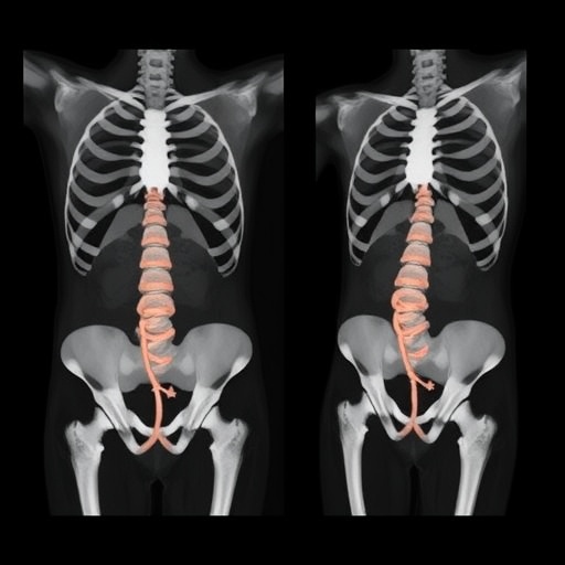
Chronic pelvic pain affects millions of people and can persist despite medication, physical therapy, nerve stimulation and surgery. A new hypothesis published in Exploratory Research and Hypothesis in Medicine proposes an unconventional route to one of the body’s key pain-processing networks: a posterior, transosseous pathway through the S1 pedicle, a dense segment of bone at the base of the spine. The concept aims to reach the superior hypogastric plexus, a network of sympathetic nerves located in the presacral and retroperitoneal region, by advancing through a fixed bony corridor rather than navigating variable soft-tissue anatomy.
The superior hypogastric plexus is already an established target for nerve blocks used in people with refractory pelvic pain, including pain associated with gynecological, urological, gastrointestinal and oncological conditions. Conventional approaches generally reach the plexus from the front or through posterior soft tissues, often relying on fluoroscopy, computed tomography or ultrasound to guide a needle around organs, vessels and other sensitive structures. Although these techniques can provide meaningful relief, their accuracy may be affected by body habitus, anatomical variation, operator experience and the position of nearby blood vessels. The newly proposed approach seeks to replace some of that uncertainty with the reproducible architecture of the sacrum.
In the hypothesis described by João Antonio Matheus Guimarães and colleagues, a needle or specialized instrument would enter from the posterior aspect of the sacrum and follow the S1 pedicle toward the anterior sacral cortex. The S1 pedicles are routinely used by spine surgeons as entry points for instrumentation, making their orientation and morphology familiar within spinal procedures. The proposed trajectory would converge toward the sacral promontory and the presacral space, where the superior hypogastric plexus is anatomically located. The underlying idea is that a controlled passage through bone could provide a stable, predictable route to the nerve network while limiting the variability encountered when instruments travel entirely through soft tissue.
The authors describe the concept as anatomically plausible rather than clinically established. Cadaveric fluoroscopic observations reportedly supported the general direction of the pathway, with a straight course from the posterior S1 region toward the anterior sacral cortex and a pattern of contrast spread considered compatible with the area of the plexus. However, contrast distribution on imaging cannot by itself prove that the nerves were reached, nor can it demonstrate that the trajectory is safe. The study therefore does not present a treatment trial, a validated procedure or evidence that patients could benefit from the technique. Instead, it presents a testable proposal intended to stimulate anatomical and procedural research.
The most important safety issue lies at the front of the sacrum. The anterior sacral cortex separates the osseous pathway from the presacral space, but breaching that cortex could bring the instrument close to major vascular structures, including the iliac vessels, as well as the ureters, bowel and other retroperitoneal tissues. A small deviation in trajectory, an unexpected anatomical variation or uncontrolled advancement could produce severe bleeding, visceral injury, infection or neurological complications. The schematic accompanying the proposal highlights this unresolved risk by showing the converging right and left pathways and their proximity to vessels represented in the presacral region. Any future investigation would therefore need to define safe depth limits and three-dimensional relationships before human use could be considered.
The proposal also reflects a broader trend in interventional medicine: adapting established surgical corridors for new targets. Transosseous access is already central to many spinal procedures because bone can provide a rigid reference point that is less affected by soft-tissue movement or body composition. In theory, the S1 pedicle could offer similar advantages for pain interventions. A fixed corridor might reduce the variability caused by differences in pelvic anatomy and make the trajectory easier to reproduce between operators. It could also create a common technical language between spine surgeons, interventional radiologists and pain specialists. Yet familiarity with pedicle instrumentation does not automatically translate into safe access to the presacral space, where the consequences of anterior cortical penetration are fundamentally different from those encountered during routine spinal fixation.
The authors contrast the proposed route with other methods used to target the superior hypogastric plexus. Posteromedian transdiscal approaches can provide access through the intervertebral disc but may expose the disc to injury or degeneration. Ultrasound-assisted procedures can improve visualization and reduce reliance on landmarks, although image quality and interpretation remain operator-dependent. An S1 transosseous pathway could theoretically avoid some of these limitations by using the sacrum itself as a stable anatomical guide. At the same time, the technique would introduce new requirements, including precise trajectory planning, detailed assessment of the sacral pedicles and real-time awareness of the anterior cortical boundary. The potential advantage is therefore inseparable from the potential hazard.
To determine whether the idea is more than an imaging concept, the researchers propose a staged validation program. The first step would involve cadaveric dissection and direct measurement of the distances between the S1 pedicles, the anterior sacral cortex, the superior hypogastric plexus and neighboring arteries, veins, ureters and bowel. These studies could reveal whether the plexus is consistently close enough to be reached and whether a safe osseous corridor exists across different pelvic shapes. The next phase would use high-resolution computed tomography and three-dimensional reconstructions to map anatomical variability in living subjects. Such imaging could help identify patients whose vascular anatomy or sacral morphology places them at especially high risk.
Only after anatomical and imaging studies established a defensible safety profile would a clinical feasibility study be appropriate. That research would need to compare the proposed route with standard superior hypogastric plexus block techniques, focusing first on technical success, needle control, contrast behavior and complications rather than pain relief alone. Investigators would also need to determine whether the target is actually the plexus or merely an adjacent presacral compartment. Objective outcomes could include imaging-confirmed spread, changes in pain intensity and function, duration of analgesia, medication use and adverse events. Independent safety monitoring and strict stopping rules would be essential because a rare but catastrophic vascular injury could outweigh modest improvements in procedural consistency.
For now, the posterior transosseous S1 pedicle approach remains a hypothesis, not a procedure ready for routine clinical practice. Its appeal comes from a clear technical premise: a reproducible bony corridor might provide better spatial control than a variable soft-tissue route to a difficult nerve target. Its limitation is equally clear: the proposed endpoint lies beside critical retroperitoneal anatomy, and the available observations do not yet establish a safe margin. The publication’s significance is therefore less about announcing a new treatment than about defining a research question at the intersection of pain medicine and spine surgery. If future cadaveric, imaging and clinical studies confirm both access and safety, the concept could eventually expand the options for patients with otherwise intractable pelvic pain. Until then, the authors emphasize that the pathway should be regarded as experimental and should not be attempted outside carefully controlled research.
Subject of Research: A proposed posterior transosseous S1 pedicle approach for accessing the superior hypogastric plexus in the treatment of chronic pelvic pain.
Article Title: A Posterior Transosseous S1 Pedicle Approach for Accessing the Superior Hypogastric Plexus: A Hypothesis
News Publication Date: 16-Jun-2026
Web References: Exploratory Research and Hypothesis in Medicine: https://www.xiahepublishing.com/journal/erhm ; DOI: https://doi.org/10.14218/ERHM.2026.00006
References: Guimarães, João Antonio Matheus, et al. “A Posterior Transosseous S1 Pedicle Approach for Accessing the Superior Hypogastric Plexus: A Hypothesis.” Exploratory Research and Hypothesis in Medicine. DOI: 10.14218/ERHM.2026.00006
Image Credits: João Antonio Matheus Guimarães
Keywords: chronic pelvic pain, superior hypogastric plexus, S1 pedicle, transosseous approach, sacrum, pain intervention, spine surgery, presacral space, anatomical research, fluoroscopy
Tags: alternative pelvic pain interventionimaging-guided nerve proceduresinnovative pain relief strategiesminimally invasive nerve targetingnerve stimulation for pelvic painpelvic pain managementposterior transosseous S1 pedicle approachrefractory pelvic pain treatmentretroperitoneal nerve block techniquessacral bone anatomysuperior hypogastric plexus nerve blocktransosseous nerve access