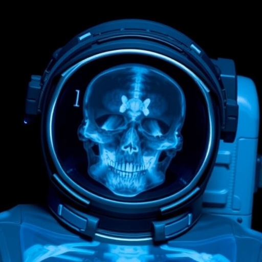
A crew aboard a commercial spacecraft has produced the first truly diagnostic X-ray images during an orbital mission, a milestone for space medicine that has long relied almost entirely on ultrasound. The results appear in Radiology, the journal of the Radiological Society of North America (RSNA), and outline how a compact, commercially available radiography system could extend medical capabilities beyond what ultrasound can deliver.
The study matters because future missions are expected to last longer and travel farther, increasing the likelihood of injuries and illnesses in orbit. Ultrasound can be helpful, but it demands substantial operator training and depends on sound-wave transmission through appropriate tissue. In contrast, X-rays are fast and provide high-value anatomical information when image quality is sufficient.
Traditional X-ray units are typically bulky, radiation-intensive, and prone to motion blur—problems intensified by the constant movement of microgravity. The research team therefore tested whether a portable system, designed for everyday use on Earth, could survive spacecraft prelaunch conditions and deliver interpretable images in space.
Prior to the Fram2 flight, the crew received four hours of training on the portable radiography workflow. Preflight imaging included hands, forearms, abdomen, pelvis, and chest X-rays, establishing baselines for comparison. The onboard setup also used an ultraportable wireless digital generator, enabling imaging with minimal medical expertise.
Fram2 launched on March 31, 2025, on a SpaceX Falcon 9 rocket into a 90-degree orbit roughly 425 to 450 kilometers above sea level. Over 3 days and 14 hours, the team acquired in-flight images of both a calibration phantom and several human anatomical regions, transmitting results to an onboard computer for immediate review by the crew.
After landing and recovery, the researchers captured postflight images designed to replicate the in-orbit protocol. Although the generator sustained superficial structural damage during touchdown, internal components and X-ray output remained functional, allowing the team to complete comparable imaging.
Three independent radiologists assessed image quality, spatial and contrast resolution, and positioning. Overall diagnostic performance was maintained between preflight and inflight acquisitions, with the main weakness appearing in the positioning scores for certain central-body images such as the chest, pelvis, and abdomen. Still, the inflight scans reached a diagnostic level for all examined regions.
Importantly, crew members reported that the system was easy to use and the protocol straightforward to follow. Estimated radiation exposure for the crew was no higher than standard clinical imaging on Earth, supporting the potential for routine onboard implementation.
Beyond human health, the study suggests X-rays could support mission-critical nonmedical tasks, from inspecting electronics and spacesuits to imaging malfunctioning satellites and analyzing lunar rover targets. The researchers argue that smaller, more rugged autonomous radiography systems could transform both space operations and broader global health logistics.
Subject of Research: People
Article Title: SpaceXray: Feasibility and Diagnostic Capabilities of On-Orbit Medical Radiography
News Publication Date: 14-Jul-2026
Web References: https://pubs.rsna.org/journal/radiology
References: Radiological Society of North America (RSNA) / Radiology
Image Credits: Radiological Society of North America (RSNA)
Keywords: Space flight; Medical imaging; Radiography; Astronauts
Tags: advancements in space medical technologyastronaut healthcare diagnosticscomparison of ultrasound and X-ray in spacediagnostic capabilities in space missionsinnovative medical tools for astronautslong-duration spaceflight health monitoringmicrogravity imaging challengesorbital X-ray imagingportable radiography systemsradiation safety in spaceradiology in microgravity environmentsSpace medicine