In the ongoing battle against breast cancer, technology often becomes the frontline defense in early detection and treatment monitoring. While mammography has long been the standard for breast cancer screening, its limitations—particularly for women with dense breast tissue and those at high risk—have spurred researchers to explore innovative imaging techniques. A team of scientists at the Massachusetts Institute of Technology (MIT) has recently made a groundbreaking advance with a portable, operator-independent 3D ultrasound system designed to facilitate more frequent and precise breast imaging. This new technology promises not only to transform cancer detection but also to enhance post-treatment surveillance in ways that were previously unimaginable.
Current ultrasound devices, although useful in follow-up diagnostics for mammogram abnormalities, come with significant constraints. They are typically bulky, expensive, and require highly trained technicians to operate effectively. This restricts their use to clinical environments, limiting accessibility, and reducing the possibility of frequent monitoring. Moreover, mammograms, the conventional screening method, must be performed annually, leaving a dangerous window during which aggressive cancers can develop undetected. These so-called “interval cancers” account for up to 30 percent of breast cancer cases and are often diagnosed at more advanced stages.
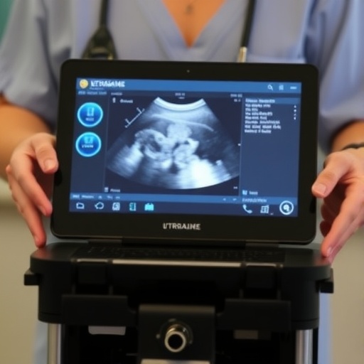
Motivated by these challenges, MIT researchers, led by Professor Canan Dagdeviren, have engineered a compact ultrasound probe coupled with an acquisition and processing unit that is approximately the size of a smartphone. Unlike traditional bulky ultrasound systems, this portable device supports fast, three-dimensional imaging of the entire breast by scanning a mere two to three locations. This spatial efficiency is a significant leap forward, potentially allowing for frequent, if not daily, breast tissue analysis outside conventional hospital settings. The ability to conduct routine screenings at home or in primary care offices could lead to the earlier identification of suspicious lesions, thereby increasing survival rates.
A central innovation in this system lies in the addition of a specialized “backing layer” behind the ultrasound transducer elements. This layer plays a crucial role in shaping the acoustic properties of the probe. It limits the dispersion of the ultrasound waves, focusing them into narrower beams that penetrate the tissue more effectively. This targeted energy transmission enhances image resolution and contrast, allowing for clearer visualization of fine anatomical structures such as cysts, microcalcifications, and small tumors. The backing layer also broadens the frequency range that the probe can operate within, amplifying its sensitivity to subtle tissue differences. Noise reduction—both acoustical and electrical—is another critical outcome, which reduces artifacts that often confound traditional ultrasound images.
Beyond hardware enhancements, the team developed an adaptive beamforming algorithm to correct for the variability in the speed of sound through different tissue types. Sound velocity is known to fluctuate between skin, fat, glandular tissue, and tumors, potentially blurring images or causing spatial inaccuracies in ultrasound scans. The novel algorithm dynamically estimates local sound speed variations and recalibrates the focusing of the ultrasound beams accordingly. This approach markedly sharpens anatomical details, achieving up to a 10 percent improvement in spatial resolution. By tailoring image formation on a tissue-by-tissue basis, the system produces highly accurate volumetric maps of breast tissue, overcoming one of the longstanding technical hurdles in ultrasound imaging.
Usability was also a primary concern in the system’s development. To democratize breast ultrasound scanning, researchers integrated a user-friendly interface paired with real-time computer vision guidance. The interface instructs users on precisely how to position the probe across successive scans, ensuring imaging consistency and reproducibility over time—a feature essential for monitoring disease progression or response to therapy. Remarkably, tests with volunteers untrained in ultrasound demonstrated that they could reliably locate and scan specific targets embedded in tissue-mimicking phantoms, outperforming conventional ultrasound scans by enabling more consistent and high-fidelity image acquisition.
The interface’s intuitive design utilizes on-screen prompts and live image feedback to minimize the learning curve, empowering patients or primary care providers to perform comprehensive breast scans safely. This capability holds immense potential for remote or resource-limited settings, where access to specialized sonographers is scarce. Conceivably, a patient could conduct self-scans at home and transmit images to clinicians for expert review, facilitating continuous disease surveillance without frequent hospital visits.
Beyond its impact on early diagnosis, the researchers envision this portable ultrasound platform playing a significant role in longitudinal disease management. For patients undergoing neoadjuvant therapy or those with known benign breast conditions like fibroadenomas or microcalcifications, routine ultrasound imaging is invaluable for tracking tissue changes over time. By enabling identical probe positioning at each scan, the system ensures that any tissue alterations are detected with higher precision, offering clinicians reliable metrics to adjust treatment plans dynamically.
Looking forward, the MIT team aims to miniaturize the system further and integrate the user interface onto smartphones or tablets. Such integration would not only increase portability but also leverage existing telemedicine infrastructure for instantaneous clinical consultation. The versatility of the underlying technology extends beyond breast cancer; it holds promise for imaging various soft tissues, including applications in ovarian cancer detection, endometriosis monitoring, and fetal health assessments. The researchers are actively pursuing commercial development avenues to bring this transformational technology to market, aspiring to make high-quality ultrasound imaging universally accessible.
This breakthrough in portable, high-resolution ultrasound imaging represents a pivotal shift in medical diagnostics, bridging technological innovation with patient-centric care. By combining sophisticated acoustic engineering, advanced signal processing algorithms, and human-centered design, the MIT research team has crafted a solution poised to revolutionize breast cancer detection and monitoring—a development with profound implications for global health.
Subject of Research: People
Article Title: Portable, real-time 3D ultrasound for operator-independent breast imaging
News Publication Date: 1-Jul-2026
Web References: http://dx.doi.org/10.1038/s41467-026-74708-3
References: Nature Communications, 10.1038/s41467-026-74708-3
Keywords: Breast cancer, Ultrasound, Portable ultrasound, 3D imaging, Medical technology, Biomedical engineering, Cancer detection, Operator-independent imaging, Acoustic beamforming, Medical equipment
Tags: accessible breast cancer diagnosticsaffordable breast cancer screening toolsbreast cancer early detection technologybreast cancer imaging technology advancementsbreast cancer interval cancer detectionbreast imaging for dense breast tissuefrequent breast cancer monitoringinnovative breast ultrasound deviceslimitations of mammography screeningoperator-independent 3D ultrasound imagingportable breast ultrasound systempost-treatment breast cancer surveillance