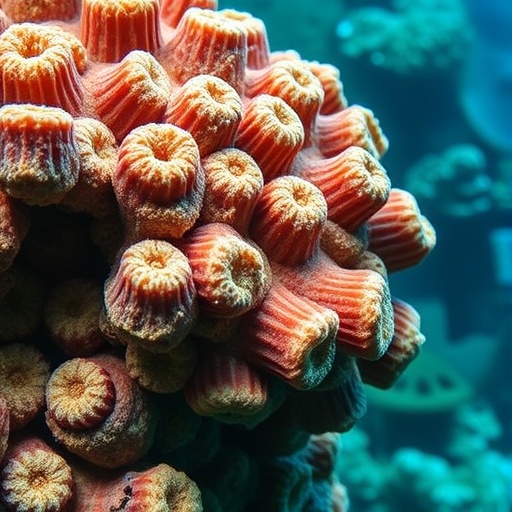
Coral reefs, often described as the rainforests of the sea, are among the most biologically diverse and valuable ecosystems on Earth. Yet, the reefs off the coast of Florida face unprecedented threats from a suite of rapidly spreading diseases, notably Stony Coral Tissue Loss Disease (SCTLD). This lethal affliction disfigures and decimates colonies at alarming rates, prompting an urgent need for advanced diagnostic methods to understand not only the visible impacts but also the hidden structural changes within coral skeletons. A team of researchers at Florida Atlantic University (FAU) has pioneered a cutting-edge approach combining micro-computed tomography (micro-CT) imaging and deep learning algorithms to delve into the complex architecture of affected coral skeletons, yielding insights with remarkable precision.
Traditional methods of assessing coral health largely rely on surface observations—visible signs of bleaching, tissue loss, or discoloration. However, these external symptoms provide limited information about the internal integrity of coral skeletons, which are crucial to the survival of reef ecosystems. The FAU research team has employed micro-CT scanning, a tomographic imaging technique that generates high-resolution, three-dimensional representations of coral skeletal structures. This non-destructive imaging modality reveals minute morphological details such as porosity variations, density fluctuations, and changes in skeletal thickness that are imperceptible through conventional methods.
By harnessing the power of deep learning—a subset of artificial intelligence capable of pattern recognition and classification—the researchers trained algorithms to interpret the voluminous data produced by micro-CT scans. The interplay between advanced imaging and AI enabled the classification of coral samples with an astonishing accuracy rate of 98%, distinguishing diseased specimens from healthy ones with minimal false positives or negatives. This fusion of technologies represents a quantum leap in our capacity to detect subtle skeletal degradation, potentially before macroscopic disease symptoms become evident.
At the heart of this innovative technique lies the capacity to quantify skeletal porosity, a key indicator of structural weakening in corals. Porosity pertains to the void spaces within the calcium carbonate matrix of the skeleton; increases in porosity can signal compromised calcification processes or bioerosion, phenomena often exacerbated by disease or environmental stresses. The FAU study quantitatively characterized the spatial distribution of pore sizes and their connectivity, providing a nuanced understanding of how SCTLD and perhaps other pathogens alter the three-dimensional framework that underpins reef resilience.
Changes in skeletal density emerged as another telling metric. Healthy coral skeletons display a uniform density profile, facilitating robustness against physical perturbations such as wave action or predator attacks. Conversely, disease-affected corals exhibited localized density reductions, signifying potential mineral dissolution or inhibited biomineralization. The 3D visualization capabilities of micro-CT allowed for mapping these density gradients across the entire coral fragment, thereby paving the way for comprehensive assessment protocols that transcend qualitative visual inspections.
Skeletal thickness, integral to the coral’s defensive armature, was also systematically measured and found to be thinner in diseased samples. This parameter is critical because it influences not only the coral’s mechanical strength but also its energy allocation for repair and growth. The precise quantification of thinning patterns not only validated disease impact hypotheses but also facilitated predictive modeling of colony survival trajectories under ongoing biological assault.
Moreover, the integration of AI expedited data processing from what would traditionally require extensive manual annotation and subjective analysis. By automating feature extraction and pattern recognition within complex volumetric data, deep learning algorithms reduced human error and variability, enabling scalable assessments across large datasets. The resultant computational pipeline offers a standardized framework that can be adapted to monitor reefs globally as they confront multifaceted threats from climate change, pollution, and emerging diseases.
One of the groundbreaking implications of this study is the potential for early disease detection. Since structural changes in the skeleton precede visible tissue loss, micro-CT combined with AI could serve as an early warning system, alerting conservationists to incipient reef degradation. This shift from reactive to proactive management heralds new possibilities in reef preservation strategies, potentially allowing targeted interventions before widespread mortality occurs.
Beyond disease monitoring, this methodology holds promise for assessing the impacts of ocean acidification and warming waters—other insidious forces driving reef decline. Both factors influence calcification rates and skeletal integrity, and subtle microstructural alterations could now be quantified with unprecedented resolution. Thus, the FAU research not only addresses immediate disease challenges but also equips the scientific community with versatile tools to quantify ecosystem responses to environmental stressors.
This research underscores the importance of interdisciplinary collaboration, merging marine biology, materials science, imaging physics, and computational analytics. Such synergy is crucial for developing holistic understanding and solutions to complex ecological crises. The marriage of micro-CT imaging with AI-driven analysis exemplifies how technology can transform environmental science, making it more precise, predictive, and scalable.
Moreover, the implications extend beyond academic insights. Policymakers, reef managers, and conservation NGOs can leverage this approach to allocate resources more effectively, prioritize vulnerable reef sections, and evaluate the success of restoration efforts. Improved structural metrics facilitate better risk assessment models and more informed decisions to safeguard marine biodiversity hotspots.
The work by FAU researchers opens a pathway toward more resilient reef ecosystems through enhanced monitoring and management. As coral diseases proliferate in frequency and severity, tools that deliver rapid, accurate assessments will be indispensable. This study provides a foundational platform upon which future technological innovations and ecological applications can be built, fostering hope for reefs amid mounting environmental uncertainty.
In a broader ecological context, the health of coral skeletons directly influences habitat complexity, which supports myriad marine species. By unveiling the hidden deterioration within coral frameworks, the FAU team illuminates a critical aspect of reef degradation that could otherwise go undetected until irreversible damage occurs. This knowledge enhances understanding of ecosystem dynamics and the cascading effects of coral diseases on marine biodiversity and fisheries.
Ultimately, the integration of micro-CT imaging and deep learning stands as a beacon of innovation in marine science, exemplifying how emerging technologies can empower conservation in the Anthropocene. As reefs confront unprecedented threats, such pioneering research is essential not only for documenting changes but for fostering action to preserve these underwater wonders for future generations.
Subject of Research: Structural impacts of Stony Coral Tissue Loss Disease on Florida’s coral reefs using micro-CT imaging and deep learning
Article Title: Advanced 3D Imaging and AI Unlock Hidden Damage in Diseased Florida Coral Reefs
News Publication Date: Not provided
Web References: Not provided
References: Not provided
Image Credits: Florida Atlantic University (FAU)
Keywords: Coral reefs, Stony Coral Tissue Loss Disease, micro-CT imaging, deep learning, coral skeleton porosity, skeletal density, skeletal thickness, reef health assessment, marine conservation, AI in ecology, coral disease diagnostics, Florida coral reefs
Tags: 3D coral imaging technologyadvanced coral disease monitoringAI in coral disease detectioncoral reef conservation technologycoral reef disease diagnosticsdeep learning for marine biologyFlorida coral reef ecosystem healthhidden skeletal damage in coralsmicro-computed tomography for coral healthnon-destructive coral assessment methodsStony Coral Tissue Loss Disease analysisstructural changes in coral skeletons