In a groundbreaking advancement for cancer diagnostics, researchers at the Institute of Science Tokyo have definitively demonstrated that small extracellular vesicles (sEVs) originating from distant tumors can traverse the bloodstream and kidney filtration barriers to be excreted intact in urine. This pivotal finding challenges long-standing assumptions about the kidney’s selective filtration and has significant implications for the future of noninvasive liquid biopsy techniques, particularly those utilizing urine as a diagnostic medium.
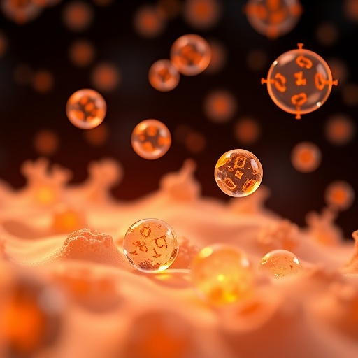
Liquid biopsy has emerged over recent decades as a transformative tool in oncology, allowing for tumor detection and monitoring through minimally invasive sampling of bodily fluids like blood and urine. Central to the promise of this approach are small extracellular vesicles—nanoscale, membrane-enclosed particles shed ubiquitously by cells, which carry molecular cargo such as proteins, lipids, and RNAs reflective of their parental cell’s state. Due to their stability and rich molecular content, sEVs circulating in biofluids are increasingly recognized as robust biomarkers for various diseases, including cancer.
Despite the theoretical potential of urine-based liquid biopsies to detect cancers even when the tumors are located far from the urinary tract, a fundamental mechanistic question persisted: how do intact tumor-derived sEVs, typically measuring between 30 and 200 nanometers in diameter, navigate the highly selective glomerular filtration barrier of the kidney? Conventional wisdom dictated that this biological filter blocks particles larger than approximately 8 nanometers, effectively precluding the passage of intact vesicles of such size into urine. This incongruity had prompted skepticism among clinicians and researchers regarding the feasibility of urinary sEV-based cancer diagnostics.
Challenging this paradigm, a collaborative research team led by Professor Takao Yasui and Associate Professor Ryosuke Kojima employed innovative molecular tracking strategies in rodent models implanted with brain, lung, and pancreatic tumors. By developing two distinct yet complementary labeling systems, the team was able to trace the biodistribution of tumor-derived sEVs with unprecedented fidelity and detail. The first system bioengineered glioma cells to produce sEVs encapsulating a synthetic RNA tracer, enabling highly sensitive molecular quantification. The second approach tagged vesicles from lung and pancreatic tumor cells with dual luminescent and fluorescent reporter proteins, allowing visualization and quantitation through advanced microscopy.
These sophisticated methodologies unambiguously substantiated the presence of tumor-derived sEVs in the urine of the animal models. Remarkably, quantitative analyses revealed a consistently higher signal intensity from tumor vesicles in urine compared to blood plasma across all tested cancer types. This counterintuitive result underscores the kidney not merely as a passive physical barrier but rather as an active participant in vesicle handling and processing within the circulatory system.
Further investigations utilized in vitro cell cultures alongside a microfluidic “glomerulus-on-a-chip” device, replicating kidney filtration dynamics under physiologically relevant flow conditions. These experiments illuminated a sophisticated mechanism whereby specialized glomerular cells engage in transcytosis—a receptor-mediated process facilitating the uptake and vesicular transport of circulating sEVs across the filtration membrane. During this transcellular journey, vesicles undergo notable alterations in size and surface protein composition, suggesting selective remodeling as part of their renal transit.
This mechanistic insight overturns archaic models that envisaged kidney filtration as exclusively size-restrictive, positioning the glomerulus instead as a dynamic regulatory interface capable of modulating extracellular vesicle profiles. The ability of tumors to shed vesicles that subsequently reach the urinary compartment intact paves the way for high-fidelity liquid biopsies tailored to noninvasive detection, monitoring, and potentially therapeutic stratification in oncology.
The research, published in the esteemed journal Science Advances, provides the first direct empirical evidence affirming that tumor-derived sEVs can be recovered from urine, including when originating from cancers located remotely, such as within the central nervous system. This breakthrough holds promise for revolutionizing early cancer detection protocols, empowering clinicians with tools to identify malignancies earlier and more accurately through simple urine tests rather than invasive biopsies or bloodstream sampling.
By situating urinary sEVs as viable, renewable biomarkers, this study lays critical technical groundwork for forthcoming diagnostic platforms that may integrate molecular tagging and nanoscale visualization. The results also prompt new inquiries into the biological functions of sEV transcytosis in kidney physiology and its broader implications in disease pathogenesis and systemic communication networks.
Professor Yasui emphasizes that “our data redefine the kidney glomerulus as an active modulator of circulating tumor vesicles, rather than a mere barrier, thereby opening transformative avenues for urinary liquid biopsy approaches.” The findings represent a compelling shift towards leveraging the body’s own biological processes for precision medicine applications.
As ongoing research continues to unravel extracellular vesicle biology, diagnostic science is poised to benefit immensely from the incorporation of urinary sEV profiling—offering a window into tumor biology that is accessible, noninvasive, and highly informative. This work exemplifies the power of interdisciplinary innovation, combining molecular biology, bioengineering, and microfluidics to push the frontiers of cancer detection.
The implications extend beyond cancer, suggesting potential utility in detecting other systemic diseases characterized by distinct sEV signatures. Ultimately, transforming urine into a diagnostic goldmine could fundamentally alter patient care paradigms, reduce healthcare costs, and improve outcomes by enabling earlier intervention.
Subject of Research: Animals
Article Title: Glomerular routing of tumor-derived extracellular vesicles substantiates urinary biopsy
News Publication Date: 20-Feb-2026
Web References: https://www.science.org/doi/10.1126/sciadv.aeb0555
Image Credits: Institute of Science Tokyo
Keywords: Cancer cells, Circulating tumor cells, Extracellular vesicles, Liquid biopsy, Kidney, Glomerular filtration barrier, Urinary diagnostics, Small extracellular vesicles (sEVs), Transcytosis, Molecular tagging, Noninvasive cancer detection, Tumor biomarkers
Tags: blood to urine vesicle transportcancer cell-derived extracellular vesiclesextracellular vesicles as cancer biomarkerskidney filtration of extracellular vesiclesliquid biopsy for distant tumorsmolecular cargo of sEVsnanoscale vesicles in oncologynoninvasive urine liquid biopsysmall extracellular vesicles in cancer diagnosticsstability of extracellular vesicles in biofluidstumor-derived sEVs in urineurine-based cancer detection methods
