Raman hyperspectral imaging has long stood at the frontier of analytical techniques capable of revealing the intricate chemical and physical characteristics of materials. By integrating vibrational spectroscopy with spatial imaging, this method provides unprecedented insight into the molecular composition and structure of a wide array of samples, from biological tissues to complex pharmaceuticals. However, a persistent challenge has been the inherently weak Raman scattering signal, which typically compels researchers to rely on lengthy acquisition times or use high-power lasers to obtain data of reasonable quality. This limitation not only slows experimental throughput but also raises concerns about sample damage and instrument cost, creating a bottleneck that has hindered broad adoption and practical deployment in many critical applications.
Addressing this fundamental barrier, a team of researchers led by Chen, Wang, and colleagues has introduced a groundbreaking computational framework known as self-optimized spectral distance (SSD). This novel approach promises to redefine the capabilities of Raman hyperspectral imaging by enabling high-quality reconstructions from data acquired under severely constrained conditions. Remarkably, SSD operates effectively with noisy measurements produced by ultra-short integration times or low-power laser excitation, thereby circumventing the traditional trade-offs between signal quality, acquisition speed, and sample viability. The implications for this advance are profound, signaling a step-change in how Raman imaging can be applied in real-world scenarios.
At its core, SSD leverages unsupervised learning principles to extract meaningful spectral information from data that would be otherwise considered insufficient for reliable analysis. Unlike deep learning methods that require extensive training datasets and computational resources, SSD independently optimizes the spectral distance metrics directly from the low-quality measurements. This self-optimization allows it to discern subtle variations in spectral fingerprints, thus reconstructing vivid Raman images that maintain both spatial and chemical fidelity. The elegance of this approach lies in its autonomy and adaptability, which permit high-throughput imaging without prior knowledge or sample labeling.
This breakthrough is particularly critical in domains where rapid, non-destructive chemical characterization is paramount. For example, in cellular biology, the ability to quickly image sub-cellular structures without prolonged exposure to intense laser beams can enable dynamic studies of living samples under physiological conditions. SSD’s capability to minimize excitation power and acquisition time could transform how researchers observe cellular processes, monitoring molecular changes in real time while preserving cell viability. Consequently, new windows may open for investigating disease mechanisms, drug responses, and cellular heterogeneity with unprecedented resolution.
Another arena poised to benefit enormously from SSD is microparticle detection. Particles at the micron scale are omnipresent in environmental, industrial, and biomedical contexts. The precision and speed offered by self-optimized spectral distance empower the rapid identification and classification of these particles, even when data quality is compromised due to limited signal strength. This means that large sample volumes can be screened quickly and reliably, facilitating applications ranging from pollution monitoring to quality control in manufacturing. The SSD methodology thus ushers in a new era of environmental and industrial Raman sensing where robustness and speed are no longer mutually exclusive.
Pharmaceutical quality control also stands to gain from this technological leap. Identifying and quantifying chemical ingredients with high spatial resolution is essential to ensure drug safety and efficacy. Conventional Raman imaging often suffers from bottlenecks in acquisition speed, particularly when high sample throughput is demanded. By dramatically cutting the time and energy required to gather meaningful spectral data, SSD could transform pharmaceutical workflows. Low-power, rapid scanning ensures that sensitive formulations remain unaltered during analysis, and high-throughput capabilities allow more extensive batch testing, improving production efficiency and regulatory compliance.
The computational architecture underlying SSD is notable for its innovation and practicality. It begins by analyzing raw, noisy hyperspectral datasets to construct a self-optimized metric space that captures subtle spectral features. This adaptive metric directly reflects the intrinsic spectral similarities and differences within the sample, enabling selective enhancement of signal components tied to chemical identities. By iteratively refining the spectral distance, SSD distinguishes meaningful Raman signals from background noise, resulting in images that reveal chemical distributions without sacrificing spatial details. The unsupervised nature of this approach eliminates dependencies on curated reference spectra or machine learning model training, which can be both data-hungry and computationally intensive.
In practical terms, the researchers demonstrated the robustness of SSD across a broad spectrum of experimental conditions and sample types. Whether imaging complex cellular components, diverse microparticles, or layered pharmaceutical ingredients, the method consistently produced high-fidelity reconstructions. Crucially, these results were achieved with at least an order of magnitude reduction in both acquisition time and laser power compared to conventional approaches. Such gains not only increase experimental throughput but also reduce potential photo-induced damage to delicate samples, broadening the applicability of Raman hyperspectral imaging to fragile biological and material systems.
The authors also discuss the potential for real-time, in situ Raman imaging workflows powered by SSD. By substantially lowering the computational and hardware demands, this technology opens pathways to portable, field-deployable Raman systems capable of rapid chemical screening. This versatility is particularly valuable for applications like environmental monitoring, where on-site sample analysis is essential, or in clinical settings, where minimally invasive diagnostics require quick and reliable molecular imaging. The advent of SSD thus paves the way for Raman imaging modalities previously limited to laboratory settings to become ubiquitous tools in diverse sectors.
Moreover, the introduction of self-optimized spectral distance methodology signals an important shift in how spectroscopic data are approached. Rather than relying solely on hardware improvements, the researchers emphasize the power of intelligent algorithmic design to transcend traditional physical constraints. This synergy between hardware and software innovations exemplifies the next generation of scientific instrumentation, where adaptive computational frameworks enhance and extend experimental capabilities. This paradigm shift could inspire similar developments in other spectroscopic and imaging modalities, fostering a new class of smart analytical devices.
Beyond its immediate practical benefits, SSD also presents exciting new research opportunities in the field of machine learning-driven spectroscopy. By demonstrating that unsupervised optimization can harness latent spectral information from noisy data, this work challenges prevailing assumptions about the necessity of large labeled datasets. It prompts renewed investigation into alternative computational approaches that exploit the underlying structure of spectral data with minimal human bias. As these computational tools mature, they promise to unlock deeper insights into complex chemical systems and enable more nuanced interpretations of vibrational spectral signatures.
Overall, the development of self-optimized spectral distance represents a landmark advancement in Raman hyperspectral imaging. It reconciles long-standing contradictions in the field—specifically, the need for high sensitivity, rapid imaging, and minimal sample perturbation—through an ingenious computational strategy. This advancement strongly aligns with broader trends toward high-throughput, minimally invasive analytical techniques that deliver rich multidimensional data with practical efficiency. The demonstrated successes in biological, environmental, and pharmaceutical contexts attest to the wide-ranging impact SSD could achieve.
Looking ahead, the integration of SSD into commercial Raman instruments and imaging platforms could catalyze widespread adoption of this method. As algorithmic optimizations become more streamlined and user-friendly, researchers and practitioners from various disciplines will be empowered to conduct complex Raman imaging experiments without requiring deep expertise in data processing. Such democratization of technology will accelerate scientific discovery and quality control across many sectors, from healthcare and pharmaceuticals to environmental science and materials engineering.
In conclusion, the innovation of self-optimized spectral distance ushers Raman hyperspectral imaging into a new era characterized by unprecedented speed, sensitivity, and accessibility. By turning what was once a prohibitive limitation—the reliance on strong Raman signals—into an opportunity for computational enhancement, this technology embodies the transformative potential of marrying physics-based measurement with cutting-edge data science. The resulting leap in imaging performance foreshadows a future where Raman spectroscopy can be deployed at scale, across a multitude of challenging scenarios, reshaping the landscape of chemical analysis and molecular imaging.
Subject of Research: Raman hyperspectral imaging, computational spectroscopy, machine learning for signal reconstruction.
Article Title: Self-optimized spectral distance for low-light high-throughput Raman hyperspectral imaging.
Article References:
Chen, Y., Wang, S., Wang, Y. et al. Self-optimized spectral distance for low-light high-throughput Raman hyperspectral imaging. Nat Comput Sci (2026). https://doi.org/10.1038/s43588-026-00957-3
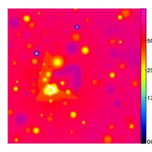
Image Credits: AI Generated
DOI: https://doi.org/10.1038/s43588-026-00957-3
Tags: advanced vibrational spectroscopy methodscomputational frameworks in spectroscopyenhancing molecular composition analysishigh-throughput Raman data acquisitionimproving Raman signal qualitylow-light Raman imaging techniqueslow-power laser Raman spectroscopyminimizing sample damage in Raman imagingRaman hyperspectral imaging challengesRaman imaging of biological tissuesself-optimized spectral distance methodultra-short integration time imaging

