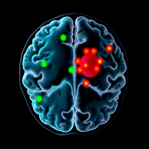
A groundbreaking advancement in artificial intelligence has emerged from the neuroscientific research community at Mass General Brigham, introducing BrainIAC—a versatile foundation model purpose-built for analyzing brain MRI data across an incredibly diverse array of medical tasks. This novel AI system transcends traditional models, which typically target singular clinical purposes, by integrating self-supervised learning techniques that enable it to understand and adapt to an extensive spectrum of neurological imaging challenges. BrainIAC’s architecture is designed to robustly extract fundamental features from unlabeled MRI datasets, thus circumventing the common bottleneck of large, meticulously annotated training data that often restricts the scalability of AI in medical imaging.
BrainIAC operates under a paradigm-shifting framework that harmonizes data heterogeneity arising from differences in imaging protocols, clinical indications, and institutional variations. Given the diversity of brain MRI scans—ranging from healthy individuals to those exhibiting complex pathologies—most existing AI models struggle to generalize results across distinct datasets and clinical contexts. By contrast, BrainIAC’s adaptable core represents a unified feature embedding space that enables the AI to perform well on tasks including but not limited to brain age estimation, molecular subtype classification of tumors, dementia risk prediction, and survival analysis for brain cancer patients, thus offering a comprehensive diagnostic platform.
Central to BrainIAC’s innovation is the utilization of self-supervised learning, a technique that leverages inherent data structures without requiring explicit supervision. This method allows the model to identify salient features intrinsic to brain MRIs by solving auxiliary tasks during pretraining. As a result, the pretrained model develops a nuanced understanding of brain anatomy and pathology that can be efficiently transferred to downstream clinical tasks with minimal labeled data. This feature is key in clinical environments where acquiring expertly annotated datasets is both expensive and time-consuming.
Extensive validation of BrainIAC’s capabilities was undertaken through a rigorous evaluation using nearly 49,000 brain MRI scans encompassing seven distinct neuroimaging applications with varying diagnostic complexity. The model demonstrated exceptional proficiency in generalizing knowledge across images of healthy brains as well as those with tumors and neurodegenerative diseases. Notably, BrainIAC excelled at conventional diagnostic tasks such as MRI sequence classification, alongside high-stakes challenges like identifying specific tumor mutation types which have critical therapeutic implications.
Comparative analyses revealed that BrainIAC significantly outperforms more narrowly focused AI frameworks, especially under conditions where training data is sparse or clinical questions are complex. This breakthrough suggests the potential for BrainIAC to be deployed effectively in real-world clinical settings that often contend with limited annotated data and diverse patient populations, thereby enhancing diagnostic precision and prognostication.
The implications of BrainIAC extend beyond improved diagnostic accuracy. By providing a unified, generalizable cognitive engine for neuroimaging analysis, it offers a promising platform for accelerating biomarker discovery at scale. Moreover, the model’s versatility enables rapid adaptation to new imaging tasks without the procedural overhead of retraining from scratch, thereby streamlining integration into existing radiological workflows and facilitating AI adoption in routine clinical practice.
From a technical standpoint, BrainIAC is built using state-of-the-art deep learning architectures tailored for volumetric imaging data. During pretraining, it harnesses multi-institutional datasets encompassing various MRI modalities to learn a rich representation of brain structure and pathology. The network’s design incorporates mechanisms to mitigate domain shifts across institutions, enabling it to maintain performance robustness when exposed to novel data sources that differ in scanner types or patient demographics.
Researchers emphasize that while BrainIAC’s performance is a significant leap forward, ongoing work is essential to extend its applicability to other neuroimaging modalities including functional MRI (fMRI) and diffusion tensor imaging (DTI). Expanding training cohorts and incorporating multimodal imaging data would further enhance the model’s predictive power, particularly for complex neurological disorders characterized by subtle, multifactorial brain changes.
The collaborative effort behind BrainIAC brought together experts in artificial intelligence, radiology, oncology, and neurology, reflecting the multidisciplinary approach required to tackle challenging clinical problems with AI. Such synergy ensures that model development is informed by deep clinical insights, thereby prioritizing relevant diagnostic endpoints and aligning AI outputs with real-world medical decision-making needs.
Mass General Brigham’s AI in Medicine (AIM) Program spearheaded this initiative, underscoring the institution’s commitment to fostering cutting-edge biomedical research that translates into tangible improvements in patient care. This aligns with their broader mission of integrating AI innovations into clinical protocols to enable precision medicine approaches tailored to individual patient profiles.
In summary, BrainIAC represents a transformative foundation model that promises to revolutionize brain MRI analysis by offering an adaptable, efficient, and clinically relevant artificial intelligence framework. Its ability to generalize across a range of neurological conditions, coupled with robustness in the face of limited training data, positions it as a pivotal resource for enhancing diagnostic workflows, predicting disease trajectories, and ultimately improving patient outcomes. As the field moves forward, BrainIAC could set a new standard for AI-powered neuroimaging, ensuring that advancements in computational modeling directly translate into enhanced healthcare delivery.
Subject of Research: People
Article Title: A Foundation Model for Generalized Brain MRI Analysis
News Publication Date: 5-Feb-2026
Web References:
https://www.massgeneralbrigham.org/
https://www.nature.com/articles/s41593-026-02202-6
References:
Tak D et al. “A foundation model for generalized brain MRI analysis” Nature Neuroscience DOI: 10.1038/s41593-026-02202-6
Image Credits: Credit: Divyanshu Tak, Mass General Brigham
Keywords:
Artificial intelligence
Cancer
Aging populations
Brain
Dementia
Tags: AI in medical imagingbrain age estimation AIbrain MRI analysisBrainIAC AI modelcancer prognosis AI toolsdisease indicators from MRIMRI data analysis across clinical contextsmultidisciplinary AI for medical diagnosticsneural imaging challengespredictive modeling in neurologyscalable AI for brain healthself-supervised learning in neuroscience

