In a groundbreaking study published in Angiogenesis, researchers have harnessed the power of 3D synchrotron X-ray imaging to illuminate the complex structure of uterine vasculature in cases of adenomyosis. This innovative technique, which employs high-energy synchrotron radiation, allows for unprecedented insights into the intricacies of blood vessels within the uterus, shedding light on a condition that has long remained shrouded in mystery. This study marks a significant advancement in the field of reproductive health and medical imaging, offering hope for more effective diagnostic and therapeutic approaches.
Adenomyosis, a condition characterized by the presence of endometrial tissue within the uterine wall, affects a substantial number of women worldwide, often leading to debilitating symptoms such as severe menstrual pain and heavy bleeding. Despite its prevalence, the underlying mechanisms of adenomyosis have not been well understood, mainly due to the challenges associated with visualizing the tissue and vascular changes that accompany the disease. Traditional imaging methods, while useful, often fall short of providing the detailed anatomical information necessary for a deeper understanding of this condition.
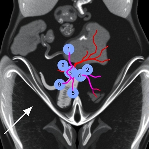
The researchers, led by Dr. V.M.W. Michels, employed advanced synchrotron X-ray imaging techniques that enable the visualization of vascular structures with high spatial resolution. This 3D approach allows for the reconstruction of complex blood vessel networks, providing insights into how vascular changes correlate with the pathophysiological features of adenomyosis. Using this state-of-the-art technology, the team was able to construct detailed 3D models of uterine vasculature, revealing previously unobserved vascular patterns associated with the condition.
What sets this study apart is the ability of synchrotron X-ray imaging to penetrate tissues without the need for traditional contrast agents, which can obscure the natural architecture of blood vessels. This non-invasive method preserves the integrity of the tissue, allowing for a more accurate representation of the uterine environment in patients with adenomyosis. By showcasing the intricacies of vascular remodeling, the study opens up new avenues for understanding how these changes might contribute to the symptoms experienced by patients.
Another pivotal aspect of the research is its implications for personalized medicine. By elucidating the unique vascular characteristics associated with adenomyosis in individual patients, healthcare providers may be able to tailor treatment strategies more effectively. For instance, identifying specific vascular markers through 3D imaging could lead to the development of targeted therapies aimed at addressing not just the symptoms but also the underlying vasculature involved in the disease process. This level of precision in treatment reflects a growing trend in modern medicine focused on individualized patient care.
Moreover, the utilization of synchrotron X-ray imaging in this context highlights the importance of interdisciplinary collaboration in advancing medical research. The convergence of physics, engineering, and medical science has enabled the development of innovative imaging technologies that not only enhance our understanding of disease mechanisms but also inform clinical practice. Such collaborations will likely become increasingly vital as the demand for advanced imaging techniques continues to grow across various branches of medicine.
The study’s findings emphasize the need for further research into the role of uterine vasculature in other gynecological conditions as well. Conditions such as endometriosis and uterine fibroids may also exhibit distinctive vascular patterns that could be elucidated through similar imaging techniques. By expanding the application of synchrotron X-ray imaging, researchers could potentially uncover common pathways underlying these disorders, leading to more comprehensive treatment options for women suffering from various reproductive health issues.
In addition to its clinical implications, this research could have broader impacts on our understanding of vascular biology. The insights gained from 3D imaging of uterine vasculature could be extrapolated to other organs and systems, paving the way for a wealth of new discoveries in vascular pathology. The ability to visualize and analyze the microvascular structures could enhance our understanding of how vascular changes affect tissue health and contribute to disease, ultimately influencing approaches to organ-specific and systemic diseases.
Looking ahead, the incorporation of advanced imaging techniques such as 3D synchrotron X-ray imaging into routine clinical practice could transform the landscape of diagnostic gynecology. As the field continues to evolve, it is essential for researchers and clinicians to work together to bridge the gap between technological innovation and patient care, ensuring that pioneering methods like these translate into tangible benefits for those affected by complex medical conditions. The excitement surrounding these advancements is palpable, as researchers envision a future where precise imaging and individualized treatment strategies fundamentally change the experience of patients.
In conclusion, the groundbreaking work by Michels and colleagues represents a significant leap forward in our understanding of adenomyosis and the complex vascular changes that accompany it. Through the application of cutting-edge synchrotron X-ray imaging, this research not only illuminates the intricacies of uterine vasculature but also lays the foundation for future explorations into gynecological conditions. As we continue to unravel the complexities of female reproductive health, it is paramount that we embrace innovative technologies that promise to enhance our diagnostic capabilities and improve clinical outcomes. The promise of precision medicine is within reach, and studies like this one serve as a beacon of hope for both researchers and patients alike.
The research not only serves as a testament to the advancements being made in imaging technologies but also underscores the need for continued investment in such interdisciplinary approaches. As science progresses and methodologies become more sophisticated, the future looks bright for improved understanding and treatment of reproductive health disorders. The ripple effects of this study will likely extend far beyond uterine health, inspiring future research in vascular biology and other fields, thereby fostering an era of discovery and improvement in patient care.
While the applications of this groundbreaking research are still emerging, its implications for women’s health cannot be overstated. By bringing to light the unseen vasculature of the uterus, Michels and her team have illuminated a path forward—one that prioritizes research-backed treatments, innovation, and ultimately, the well-being of patients. As we stand on the brink of new frontiers in medical science, the findings from this study will undoubtedly resonate within the scientific community, sparking dialogue, inspiration, and further research initiatives.
The exploration of uterine vasculature through advanced imaging techniques such as synchrotron X-ray is only the beginning of what could become a significant movement in understanding female reproductive health. As we continue to reveal the unseen, every discovery brings us one step closer to a future where conditions like adenomyosis are fully understood and effectively treated, empowering women worldwide to lead healthier lives.
Subject of Research: Uterine vasculature in adenomyosis
Article Title: Revealing the unseen: 3D synchrotron X-Ray imaging of uterine vasculature in adenomyosis
Article References: Michels, V.M.W., Szmul, A., Jacob, J. et al. Revealing the unseen: 3D synchrotron X-Ray imaging of uterine vasculature in adenomyosis. Angiogenesis 29, 3 (2026). https://doi.org/10.1007/s10456-025-10004-w
Image Credits: AI Generated
DOI: https://doi.org/10.1007/s10456-025-10004-w
Keywords: Adenomyosis, 3D imaging, synchrotron X-ray, uterine vasculature, reproductive health.
Tags: 3D imaging of uterine blood vesselsadenomyosis diagnosis advancementsendometrial tissue in uterine wallhigh-energy synchrotron radiation applicationsinnovative imaging techniques for adenomyosismedical imaging breakthroughsreproductive health researchsevere menstrual pain causessynchrotron X-ray technology in medicinetherapeutic approaches for adenomyosis.understanding adenomyosis mechanismsvisualization of uterine vasculature

