Recent advancements in the medical imaging field have paved the way for innovative approaches to abdominal vascular imaging, particularly highlighting the role of ferumoxytol, an iron oxide nanoparticle. This cutting-edge technique, as detailed by Rees et al. in their upcoming paper, is set to transform how pediatric radiologists evaluate and diagnose vascular conditions within the abdomen. With the initiation of this groundbreaking study, the authors delve into the practical applications, efficacy, and safety of ferumoxytol-enhanced imaging methodologies.
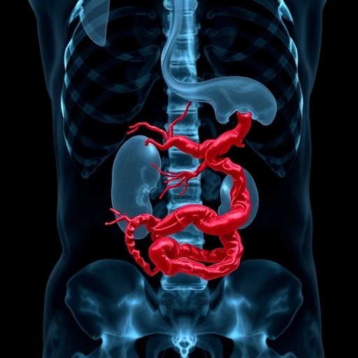
Ferumoxytol, initially approved as a treatment for anemia, has garnered attention for its imaging capabilities due to its unique properties. The nanoparticle serves as a contrast agent for magnetic resonance imaging (MRI), offering clear visualization of vascular structures under examination. The ability of ferumoxytol to remain in the vascular system for extended periods improves image quality, allowing clinicians to capture detailed snapshots of abdominal blood vessels. This aspect is particularly advantageous in pediatric populations, where conventional contrast agents may present risks.
The intricate mechanisms through which ferumoxytol operates in vascular imaging are equally fascinating. The ferumoxytol nanoparticles interact with magnetic fields during MRI scans, enhancing the contrast of blood vessels against surrounding tissues. This differential imaging capability supports the identification of various pathological conditions, including vascular anomalies, tumors, and inflammatory diseases. By employing non-invasive methods, this technique reduces the need for surgical interventions and enables higher diagnostic confidence.
One of the pivotal factors in this burgeoning field is the safety profile of ferumoxytol. While traditional contrast agents often carry risks of adverse reactions, ferumoxytol has demonstrated a strong safety record. The study conducted by Rees et al. emphasizes this aspect, noting that the incidence of hypersensitivity reactions is notably lower than that associated with iodinated contrasts. This characteristic not only positions ferumoxytol as a favorable alternative but also encourages its adoption in clinical practices.
In addition to safety, the authors provide a comprehensive analysis of the imaging protocols employed in their study. They delineate the optimal dosages and timing for administering ferumoxytol, ensuring that healthcare providers can maximize the benefit while minimizing potential drawbacks. The intricacies behind the preparation, injection, and scanning processes are discussed in depth, equipping radiologists with the necessary knowledge to implement this innovative technique effectively.
Moreover, the paper highlights comparative studies with conventional imaging techniques, underscoring the superior diagnostic capabilities of ferumoxytol-enhanced MRI. When juxtaposed with computed tomography (CT) scans and standard MRI protocols, ferumoxytol has been shown to deliver a clearer and more precise depiction of vascular structures. This comparative advantage suggests that adopting this imaging method could lead to improved patient outcomes and more accurate diagnoses in pediatric cases.
The authors also touch upon the future of ferumoxytol in expanding the horizons of abdominal imaging. As technology advances, the potential applications of this nanoparticle extend beyond mere vascular imaging. Ongoing research aims to explore how ferumoxytol could enhance imaging in oncology or other organ systems, potentially revolutionizing diagnostic imaging across multiple medical fields.
As awareness grows regarding the potential of ferumoxytol, it is crucial for medical institutions to invest in training programs and infrastructure to support its integration into routine practice. Continued collaboration between radiologists, pediatricians, and pharmaceutical developers will ensure that this promising technology receives the necessary support and validation in clinical settings, ultimately aiming to enhance diagnostic capabilities and patient care.
In conclusion, the study by Rees et al. represents a significant advancement within the realm of pediatric radiology. As the healthcare sector embraces more innovative imaging strategies, ferumoxytol stands out as a beacon of hope for enhanced abdominal vascular imaging. The findings are a testament to the evolution of medical imaging, promising a new era where non-invasive techniques can lend invaluable insights into complex pediatric conditions, optimizing patient management while alleviating procedural risks.
This synthesis of technology and medicine heralds a new chapter in contemporary radiology, encouraging clinicians to remain at the forefront of research developments. As 2026 unfolds, the rippling effects of this research will undoubtedly echo throughout the medical community, potentially reshaping the diagnostic landscape for years to come.
Subject of Research: Abdominal vascular imaging with ferumoxytol
Article Title: Abdominal vascular imaging with ferumoxytol – how we do it
Article References:
Rees, M., Salman, R., Krishnamurthy, R. et al. Abdominal vascular imaging with ferumoxytol – how we do it.
Pediatr Radiol (2026). https://doi.org/10.1007/s00247-026-06515-3
Image Credits: AI Generated
DOI: 22 January 2026
Keywords: Abdominal vascular imaging, ferumoxytol, pediatric radiology, MRI, contrast agents, safety, diagnostic imaging, vascular anomalies, non-invasive techniques.
Tags: contrast agents for MRIdiagnosing vascular conditions in childrenefficacy of ferumoxytol as a contrast agentenhanced imaging capabilities with ferumoxytolferumoxytol abdominal vascular imaginginnovative imaging methods for vascular conditionsiron oxide nanoparticles in MRImagnetic resonance imaging advancementspediatric radiology and ferumoxytolpediatric vascular imaging techniquessafety of ferumoxytol in imagingvisualization of abdominal blood vessels
