In a groundbreaking and rare case in the realm of pediatric medicine, researchers have delineated the complex and unusual presentation of a hepatic germ cell tumor with intrabiliary involvement. This study brings to light the diagnostic challenges and atypical imaging characteristics associated with such tumors, primarily through the lens of magnetic resonance imaging (MRI). The case highlights a critical intersection of oncology and radiology, showcasing how advanced imaging techniques can aid in the identification of rare pathologies.
Hepatic germ cell tumors are notably uncommon, constituting a small fraction of all pediatric tumors. They typically originate from primordial germ cells, which have the potential to develop into various tissue types. The rarity of these tumors, especially in the liver, poses unique challenges for clinicians. This case exemplifies the need for a heightened awareness among healthcare providers when faced with atypical presentations in pediatric patients. The research underscores that these tumors can manifest in ways that might initially elude even seasoned medical professionals.
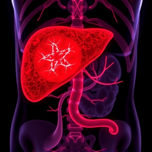
MRI plays a pivotal role in the diagnosis of hepatic tumors, providing detailed images that reveal not just the anatomy but also the pathological characteristics of the tissues in question. In this case, the MRI findings indicating intrabiliary involvement were both striking and unconventional, prompting an immediate investigation into the underlying pathology of the liver lesions. The images obtained demonstrated a complex relationship between the tumor and the biliary system, raising important questions about the management and treatment of similar cases.
Intrabiliary involvement is particularly noteworthy because it can complicate the clinical picture considerably. Tumors that invade or compress the biliary ducts can lead to cholestasis and subsequent liver dysfunction. The implications for patient management become significant, as clinicians must balance the need for surgical intervention against the potential risks associated with such procedures. The research highlights how a comprehensive understanding of the surgical anatomy is crucial for planning any operative approach to these tumors.
The findings also suggest that radiologists and oncologists must work closely together to develop a coherent diagnostic plan. Clear communication between these specialists is essential to interpret MRI findings accurately and to outline a differential diagnosis that considers all potential causes of hepatic masses. Collaboration ensures that all angles are explored and that the most effective treatment pathways are pursued.
The complexity of diagnosing hepatic germ cell tumors is echoed in the case where standard imaging techniques were initially inconclusive. As the tumor progressed and exhibited changes on the MRI, it became increasingly evident that a differential diagnosis had to be established promptly. This scenario emphasizes the importance of continuous education and familiarity with rare pathologies for radiologists and oncologists alike.
Histopathological evaluation remains crucial, despite the advancements in imaging technology. While MRI provides a wealth of information, it does not replace the need for tissue diagnosis. The correlation between imaging findings and biopsy results is vital for establishing the nature of the tumor and determining the most appropriate therapeutic intervention. This case reinforces the need for a multidisciplinary approach, bringing together expertise from radiology, pathology, and surgical oncology.
Moreover, the unique nature of this case sheds light on the potential variance in tumor behavior based on their location and biological characteristics. The intrabiliary involvement of the hepatic germ cell tumor challenged the typical presentation that most clinicians would expect, suggesting that more research is needed to fully understand the behavior of these tumors. Investigating these variables can provide insights into prognosis and guide future research efforts.
As the field of pediatric oncology continues to evolve, the treasures unearthed in rare cases such as this one emphasize the importance of clinical vigilance. Every atypical presentation might offer new knowledge and foster advancements in diagnostics and treatment protocols for rare tumors. The partnership between radiologists and oncologists can lead to better outcomes, provided that there is an effective exchange of information.
The technological advancements in magnetic resonance imaging have paved the way for increasingly sophisticated diagnostic capabilities. Radiology plays a central role in identifying the nuances of tumor presentations, and ongoing innovation will only enhance these tools further. The study advocates for investment in research and development within this domain, as breakthroughs in imaging technology could positively impact patient management.
Furthermore, this case serves as a reminder of the broader implications of rare tumors and their presentations. Understanding the intersection of genetics, pathology, and imaging in the context of hepatic germ cell tumors is paramount. Further investigation into the genetic and molecular characteristics of these tumors could prove beneficial in the long run. Identifying molecular markers may pave the way for innovative therapeutic strategies tailored to the specific vulnerabilities of these tumors.
In conclusion, the remarkable case of intrabiliary involvement of a hepatic germ cell tumor highlighted by Agarwal et al. amplifies our understanding of these rare pediatric tumors. The integration of advanced MRI imaging techniques, vigilant diagnosis, and interdisciplinary collaboration illustrates the comprehensive efforts required to navigate the challenges posed by such cases. As the investigation continues, it holds potential for significant implications in the fields of pediatric oncology and radiology, ultimately striving for improved patient outcomes.
This case study not only contributes to the existing literature but also calls for increased awareness and research investments in pediatric malignancies, particularly those that are rare and underrepresented. It is hoped that through continued advocacy and collaboration, the medical community will uncover the mysteries surrounding these challenging tumors, fostering a deeper understanding and enhancing overall care for affected children.
Subject of Research: Hepatic Germ Cell Tumor with Intrabiliary Involvement
Article Title: Intrabiliary involvement of a hepatic germ cell tumor on magnetic resonance imaging: a rare presentation
Article References:
Agarwal, A., Sharma, G. & Dwivedi, S. Intrabiliary involvement of a hepatic germ cell tumor on magnetic resonance imaging: a rare presentation.
Pediatr Radiol (2026). https://doi.org/10.1007/s00247-026-06516-2
Image Credits: AI Generated
DOI: 10.1007/s00247-026-06516-2
Keywords: Hepatic Germ Cell Tumor, Intrabiliary Involvement, MRI, Pediatric Oncology, Radiology
Tags: advanced imaging techniques in diagnosisatypical presentations in pediatric medicinediagnostic challenges in pediatric tumorshealthcare provider awareness in rare conditionsintrabiliary involvement in tumorsliver tumors in childrenMRI imaging in oncologypediatric oncology case studyprimordial germ cells and tumorsradiology in tumor identificationrare hepatic germ cell tumoruncommon pediatric cancers
