In a groundbreaking advancement in the understanding of Parkinson’s disease (PD), recent research has unveiled distinct structural and functional asymmetries in the brains of affected individuals, differentiating between the brain-first and body-first subtypes. Utilizing cutting-edge magnetic resonance imaging (MRI) techniques, this study provides unprecedented insights into the neuroanatomical and neurophysiological divergences that characterize these subtypes, hinting at underlying pathogenic mechanisms and potential diagnostic biomarkers.
The research, conducted by Shi, J., Zhang, H., Wang, X., and colleagues and published in npj Parkinson’s Disease, addresses a long-standing puzzle in neurological research: why do Parkinson’s patients often exhibit asymmetric symptoms, and how might this reflect deeper, subtype-specific brain alterations? Traditionally, Parkinson’s disease has been clinically identified by motor symptoms such as tremors, rigidity, and bradykinesia that typically manifest asymmetrically. However, the nuances of how brain structure and function mirror these clinical asymmetries remained elusive until now.
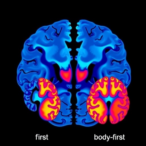
Using high-resolution MRI modalities, including both structural and functional imaging, the research team meticulously analyzed axial brain asymmetries—the differences between corresponding regions in the left and right hemispheres—among patients classified into brain-first and body-first Parkinson’s disease subtypes. The brain-first subtype is characterized by initial neurodegeneration originating within the central nervous system, whereas the body-first subtype suggests a peripheral origin with retrograde propagation toward the brain.
The investigators applied advanced neuroimaging protocols to quantify differences in cortical thickness, subcortical volume, and resting-state functional connectivity. Their findings revealed pronounced lateralization patterns in critical regions implicated in Parkinson’s pathology, such as the substantia nigra, striatum, and various cortical motor and sensory areas. Notably, the brain-first subtype exhibited more pronounced asymmetry in the substantia nigra’s structural integrity, correlating with the typically unilateral initial motor deficits observed in these patients.
Simultaneously, functional MRI analyses exposed divergent patterns of network connectivity. Brain-first patients showed disrupted coupling between the basal ganglia and motor cortex predominantly on the affected side, aligning with the hypothesis that dopaminergic neuronal loss in these areas precipitates motor symptom onset. Conversely, individuals with body-first Parkinson’s displayed more symmetric but globally altered connectivity profiles, potentially reflecting the systemic and multisite progression of alpha-synuclein pathology inherent to their subtype.
The study also delved into axial asymmetries of cortical regions beyond the classical motor circuitry. Intriguingly, sensory integration areas and limbic structures revealed subtype-specific lateralization, which may underpin the differential presentation of non-motor symptoms across brain-first and body-first patients, including variations in cognitive impairment, mood disturbances, and autonomic dysfunction. This multidimensional approach highlights the complexity and heterogeneity of Parkinson’s disease beyond mere motor symptomatology.
Methodologically, the researchers leveraged novel analytic frameworks to map and quantify asymmetries, including voxel-based morphometry (VBM) to capture subtle regional volumetric changes, and seed-based resting-state functional connectivity analyses to interrogate functional dynamics. The integration of these techniques allowed a comprehensive portrait of how structural degeneration and network dysregulation interplay within asymmetric spatial frameworks.
From a clinical perspective, the implications are profound. Recognizing the distinct axial asymmetry signatures linked to Parkinson’s subtypes could refine diagnostic accuracy, facilitating early and subtype-specific interventions. Mapping these neuroimaging biomarkers may enable clinicians to stratify patients more effectively, tailor therapeutic strategies, and monitor disease progression with enhanced sensitivity.
Moreover, this study opens avenues for exploring the mechanistic underpinnings of Parkinson’s disease asymmetry. By correlating imaging phenotypes with molecular markers and clinical data, future research can dissect how neurodegenerative cascades evolve differently in brain-first versus body-first PD. This could unravel novel targets for disease-modifying therapies aimed at halting or reversing the spread of pathology.
Importantly, the team’s findings underscore the value of precision medicine approaches in neurodegenerative diseases. The delineation of subtype-specific brain changes through reproducible and non-invasive imaging modalities promises to bridge the gap between clinical heterogeneity and underlying biological variability—a crucial step toward personalized healthcare.
The research also contributes to ongoing debates regarding the initiation and propagation of alpha-synuclein pathology in PD. The brain-first subtype’s pronounced unilateral nigral degeneration supports models where initial CNS involvement drives disease onset. Conversely, the body-first subtype’s symmetrical connectivity disruptions lend credence to peripheral origins with systemic impact. These insights refine conceptual frameworks and may guide future neuropathological and biomarker investigations.
Furthermore, the study capitalizes on sophisticated image processing pipelines, employing automated segmentation and lateralization indices to ensure objective and reproducible measurements of asymmetry. The robust statistical design controlled for confounding factors such as age, disease duration, and medication status, enhancing the validity of the observed subtype differences.
While promising, the authors acknowledge limitations including the need for longitudinal studies to capture temporal dynamics of asymmetry evolution and the necessity to validate findings across larger, multi-center cohorts. Additionally, integrating complementary biomarkers such as cerebrospinal fluid analysis and PET imaging could enrich the characterization of subtype-specific pathophysiology.
Overall, this landmark work represents a substantive leap in delineating the neuroanatomical and functional complexities inherent to Parkinson’s disease subtypes. The elucidation of axial asymmetry patterns not only deepens scientific understanding but also charts a strategic path toward improved clinical management and therapeutic innovation.
As Parkinson’s disease continues to challenge millions worldwide, harnessing the power of neuroimaging to decode its enigmatic heterogeneity is both timely and transformative. This study exemplifies the integration of advanced imaging science with clinical neurology, positioning the field to unlock targeted, effective interventions that recognize and respect the disease’s multifaceted nature.
In conclusion, the discovery of distinct MRI-based axial asymmetry profiles that discriminate brain-first from body-first Parkinson’s disease subtypes constitutes a pivotal advance. It highlights the critical role of structural and functional brain lateralization in disease manifestation and progression. This refined neuroimaging lens will be essential for future endeavors aiming to personalize care, develop novel therapeutics, and ultimately improve outcomes for patients grappling with this complex and devastating disorder.
Subject of Research: Parkinson’s Disease Subtype Differentiation via MRI-based Structural and Functional Brain Asymmetry
Article Title: MRI structural and functional axial asymmetry in the brain-first versus body-first subtypes of Parkinson’s disease
Article References:
Shi, J., Zhang, H., Wang, X. et al. MRI structural and functional axial asymmetry in the brain-first versus body-first subtypes of Parkinson’s disease. npj Parkinsons Dis. (2025). https://doi.org/10.1038/s41531-025-01219-1
Image Credits: AI Generated
Tags: advancements in Parkinson’s disease understandingasymmetrical symptoms in Parkinson’sBrain-First vs Body-First Parkinson’scentral vs peripheral origins of neurodegenerationfunctional imaging in Parkinson’s researchMRI techniques in neurodegenerative diseasesneuroanatomical differences in PDneurophysiological characteristics of PDParkinson’s disease brain asymmetryParkinson’s disease diagnostic biomarkersstructural brain alterations in Parkinson’ssubtype-specific Parkinson’s disease research


