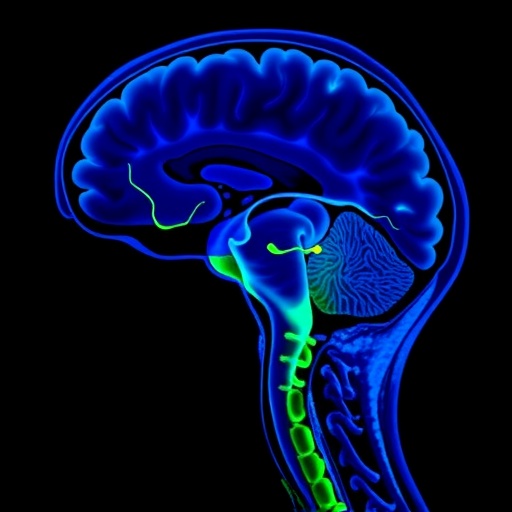
In a groundbreaking development within oncology and neuro-oncology, researchers have unveiled compelling evidence positioning hepcidin as a potent biomarker for detecting leptomeningeal metastases (LMD) in patients battling metastatic breast cancer. This discovery promises to revolutionize current diagnostic paradigms, offering hope for earlier and more accurate identification of this devastating neurological complication. LMD, characterized by the spread of cancer cells to the leptomeninges surrounding the brain and spinal cord, presents a formidable challenge due to its elusive detection and aggressive clinical course.
Leptomeningeal metastatic disease afflicts a notable fraction—ranging from 5 to 19 percent—of patients with solid tumors, with breast cancer standing prominently among the predominant primary sources. The insidious nature of LMD is compounded by the central nervous system’s (CNS) inherent defense mechanisms, notably the blood-brain barrier, which limits the penetration of conventional therapeutic agents, thereby complicating treatment and potentially contributing to the observed increase in LMD incidence as patients enjoy prolonged survival from systemic therapies.
Currently, the gold standards for diagnosing LMD rest upon cerebrospinal fluid (CSF) cytology and magnetic resonance imaging (MRI). However, these modalities are fraught with limitations; CSF cytology, while specific, often lacks sensitivity in early disease stages, and MRI may fail to reveal subtle or diffuse leptomeningeal involvement. This diagnostic gap underscores a pressing need for sensitive, reliable biomarkers capable of facilitating earlier detection and improved disease monitoring.
In an innovative study published in the journal BMC Cancer, an investigative team led by Delaby et al. embarked on an exploratory analysis aimed at identifying viable biomarkers within CSF and serum that could serve as diagnostic indicators of LMD in metastatic breast cancer patients. Their focus encompassed several neurodegeneration-associated proteins—neurofilament light chain (NfL), glial fibrillary acidic protein (GFAP), Tau protein—as well as the iron-regulating hormone hepcidin, previously unexplored in this context.
The study cohort comprised 49 adult patients, stratified into two groups: 18 individuals with confirmed LMD based on CSF cytology and 31 patients without cytological evidence of LMD. Through meticulous quantification assays, the researchers measured concentrations of the target proteins in CSF and serum samples. Remarkably, median CSF hepcidin levels were substantially elevated in LMD-positive patients (1.4 ng/mL) compared to their LMD-negative counterparts (0.3 ng/mL), a difference that was statistically significant and underscored hepcidin’s potential role as a biomarker reflective of leptomeningeal disease burden.
To quantitatively assess diagnostic performance, the researchers employed receiver operating characteristic (ROC) curve analysis, revealing that CSF hepcidin attained an area under the curve (AUC) of 0.909—indicative of excellent discriminative ability. This surpassed other measured biomarkers; proteinorachy achieved an AUC of 0.841, whereas NfL, GFAP, and total Tau demonstrated more modest but still statistically significant AUC measures ranging from approximately 0.7 to 0.72. Intriguingly, serum hepcidin did not exhibit similar diagnostic utility, with an AUC barely above 0.5, suggesting that the elevations in hepcidin are localized principally within the CSF compartment in the setting of LMD.
The implications of these findings are multifaceted. Hepcidin, traditionally recognized as the master regulator of systemic iron homeostasis, is synthesized predominantly in the liver but also in other tissues, including those within the CNS under pathophysiological conditions. Its elevation in the CSF during LMD may reflect iron metabolism dysregulation associated with tumor infiltration or secondary inflammatory responses, thereby offering a window into the microenvironmental alterations wrought by leptomeningeal metastases.
Clinicians grappling with the diagnosis of LMD currently contend with the challenges of repeat lumbar punctures and reliance on imaging that may be inconclusive. Hepcidin’s emergence as a biomarker heralds a future where a simple CSF assay might significantly boost diagnostic confidence, permitting earlier initiation of targeted therapies and potentially improving neurological outcomes. This biomarker could also serve as a valuable tool in monitoring disease progression or response to treatment, thus tailoring patient management strategies.
Furthermore, the study sheds light on the broader biological dynamics at play within the CNS during metastatic invasion. The elevated presence of neurodegenerative markers such as NfL and GFAP, albeit with less diagnostic precision than hepcidin, mirrors neuronal and glial injury associated with leptomeningeal involvement, offering complementary insights that may enrich the clinical assessment landscape.
The research, while exploratory and inviting further validation in larger cohorts, sets a precedent for the integration of molecular diagnostics into neuro-oncological practice. A nuanced understanding of the interplay between iron metabolism and cancer biology in the CNS could provoke novel therapeutic avenues, potentially exploiting hepcidin pathways to modulate disease progression or augment treatment efficacy.
In the context of metastatic breast cancer—a disease already complex due to its propensity for systemic dissemination and heterogenous molecular subtypes—this advancement in biomarker identification is particularly auspicious. With survival improvements due to breakthroughs in systemic therapies, managing CNS involvement remains a critical frontier, and enhanced diagnostic tools like CSF hepcidin measurement are essential to meet this challenge.
As the oncology community strives to refine precision medicine approaches, findings such as these reinforce the importance of biomarker-driven diagnostics. The clinical translation of CSF hepcidin evaluation could, in time, evolve into a standardized component of neuro-oncological assessment, aligning with personalized treatment paradigms that optimize outcomes and minimize neurological morbidity.
In conclusion, the study spearheaded by Delaby and colleagues presents compelling evidence positioning CSF hepcidin as an emerging, highly sensitive predictor biomarker for leptomeningeal metastases in metastatic breast cancer patients. This discovery stands to substantially enhance diagnostic accuracy, enabling earlier detection and improved therapeutic decision-making. Future research endeavors aimed at validating these findings and elucidating the underlying mechanistic pathways will be crucial in harnessing the full clinical potential of hepcidin within this challenging clinical domain.
Subject of Research:
Breast cancer-derived leptomeningeal metastases and biomarker identification via cerebrospinal fluid analysis.
Article Title:
Hepcidin as an emerging predictor biomarker of leptomeningeal metastases in patients with metastatic breast cancer
Article References:
Delaby, C., Al Herk, A., Hirtz, C. et al. Hepcidin as an emerging predictor biomarker of leptomeningeal metastases in patients with metastatic breast cancer. BMC Cancer 25, 1801 (2025). https://doi.org/10.1186/s12885-025-15124-6
Image Credits: Scienmag.com
DOI: 21 November 2025
Keywords:
Leptomeningeal metastases, metastatic breast cancer, hepcidin, cerebrospinal fluid biomarkers, diagnostic biomarkers, neurofilament light chain, glial fibrillary acidic protein, Tau protein, CNS metastasis, iron metabolism, neuro-oncology, biomarker discovery
Tags: blood-brain barrier and cancer treatmentcancer spread to the central nervous systemcerebrospinal fluid cytology limitationschallenges in detecting leptomeningeal metastasesdiagnosing leptomeningeal metastatic diseaseearly detection of leptomeningeal metastaseshepcidin as a biomarker for leptomeningeal metastasesleptomeningeal metastases in breast cancermagnetic resonance imaging for LMDmetastatic breast cancer complicationsneuro-oncology advancementstherapeutic challenges in leptomeninge


