In a groundbreaking advancement that could redefine the early diagnosis of Parkinsonism, a team of researchers have harnessed the power of advanced MRI techniques to identify biomarkers capable of predicting the development of this neurodegenerative disorder in individuals with idiopathic rapid eye movement sleep behavior disorder (iRBD). The study, recently published in npj Parkinson’s Disease, demonstrates the remarkable potential of susceptibility and diffusion MRI modalities to serve as non-invasive, predictive tools that may revolutionize how Parkinsonism is detected and managed long before clinical symptoms manifest.
Idiopathic rapid eye movement sleep behavior disorder, characterized by abnormal behaviors during the REM phase of sleep, has long been recognized as a significant prodromal marker for Parkinsonism and related synucleinopathies. However, the clinical challenge remains in stratifying which iRBD patients are most likely to progress to full-blown Parkinson’s disease or related disorders. This study elegantly addresses this issue by employing advanced neuroimaging biomarkers that quantify brain changes linked to neurodegeneration with unparalleled sensitivity.
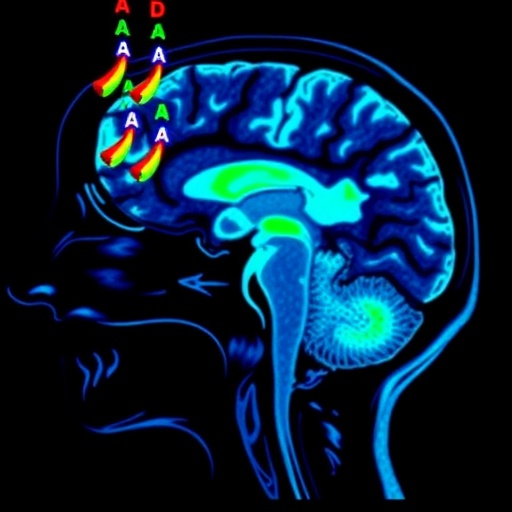
The core technological innovation lies in the application of susceptibility-weighted imaging (SWI) and diffusion magnetic resonance imaging (dMRI) to detect microstructural and iron-related alterations in strategic brain regions. SWI exploits variations in magnetic susceptibility to accentuate iron accumulation, a pathological hallmark of Parkinson’s disease, especially in the substantia nigra. Meanwhile, dMRI measures the diffusion of water molecules along neuronal pathways, revealing subtle microstructural disruptions that precede gross anatomical changes.
By integrating these imaging modalities, the researchers constructed a biomarker profile that not only distinguishes iRBD patients at risk but also quantitatively predicts the temporal trajectory toward Parkinsonism development. This predictive capability opens a promising avenue for early intervention, allowing clinicians to deploy neuroprotective therapies during a critical window when neuronal loss might still be mitigated.
A cohort of patients diagnosed with iRBD underwent comprehensive MRI screening, with follow-up clinical assessments spanning several years. The imaging data unveiled distinct patterns of increased iron deposition and disrupted diffusion metrics consistent with nigrostriatal degeneration in those who eventually manifested Parkinsonism. Notably, these biomarkers appeared well before traditional motor symptoms emerged, illustrating the power of this approach to detect subclinical disease processes.
The implications of these findings extend beyond diagnostic enrichment. The detailed characterization of pathological brain changes through MRI could also serve as objective endpoints in clinical trials testing novel therapeutics aimed at halting or slowing Parkinson’s disease progression. Researchers and pharmaceutical developers now have a quantifiable metric to assess treatment efficacy in a population at greatest risk.
Moreover, the non-invasive nature of MRI scanning ensures patient compliance and feasibility in diverse clinical settings, including longitudinal monitoring. Unlike invasive biomarker sampling or costly molecular imaging with radiotracers, susceptibility and diffusion MRI can be readily integrated into current diagnostic workflows, streamlining patient evaluation and follow-up.
This study also highlights the critical role of iron metabolism dysregulation in the pathogenesis of Parkinsonism. Iron accumulation in the substantia nigra is both a diagnostic marker and a potential contributor to oxidative stress and neuronal death. By mapping these variations in vivo with SWI, researchers provide direct evidence correlating iron burden with disease onset among vulnerable individuals.
The diffusion MRI findings complement this by exposing microstructural impairments within the nigrostriatal pathways, indicating demyelination, axonal injury, or neuronal loss at stages traditionally considered presymptomatic. These changes underscore the silent progression of neurodegeneration and emphasize the urgent need for tools capable of capturing these early signals.
The study’s multivariate approach, combining susceptibility and diffusion metrics, represents a significant leap in biomarker science. The integration enhances accuracy, reduces false positives, and delineates a more comprehensive neurodegenerative signature, critical for precise individualized risk profiling.
Furthermore, the research team’s methodological rigor, employing longitudinal designs with extensive clinical correlation, strengthens the validity of their conclusions. This robust framework sets a new standard for biomarker validation, fostering confidence in widespread clinical application and research adoption.
Importantly, the findings open pathways toward personalized medicine in Parkinsonism. By identifying high-risk individuals early, personalized intervention strategies can be developed, ranging from lifestyle modifications to pharmacological therapies, tailored to individual biomarker profiles and progression risk.
The study also prompts a reevaluation of current diagnostic criteria and encourages the integration of advanced neuroimaging markers into consensus guidelines for Parkinson’s disease and iRBD management. Such a paradigm shift will necessitate interdisciplinary collaboration between neurologists, radiologists, and sleep medicine specialists.
In sum, the convergence of susceptibility and diffusion MRI biomarkers heralds a transformative era in neurodegenerative disease research. This pioneering work not only demystifies the prodromal phase of Parkinsonism but also empowers clinicians with innovative tools to predict, monitor, and potentially alter the disease course.
Future research directions will likely explore the scalability of these imaging biomarkers in larger, more diverse populations, assess their utility alongside molecular and genetic markers, and refine imaging protocols for optimal sensitivity and specificity.
Ultimately, this landmark study paves the way for earlier diagnosis and intervention strategies that could profoundly improve patient outcomes, reduce disease burden, and foster hope for effective management of Parkinsonism and related disorders.
As the medical community continues to grapple with the complexities of neurodegenerative diseases, advances such as these illuminate a path forward, demonstrating that the fusion of cutting-edge imaging technology with clinical insight can unlock new frontiers in understanding and combating conditions like Parkinson’s disease.
Subject of Research: The identification of susceptibility and diffusion MRI biomarkers for predicting the development of Parkinsonism in patients diagnosed with idiopathic rapid eye movement sleep behavior disorder (iRBD).
Article Title: Susceptibility and diffusion MRI biomarkers predict development of Parkinsonism in iRBD.
Article References:
Varga, Z., Nepozitek, J., Hlavnicka, J. et al. Susceptibility and diffusion MRI biomarkers predict development of Parkinsonism in iRBD. npj Parkinsons Dis. 11, 332 (2025). https://doi.org/10.1038/s41531-025-01174-x
DOI: https://doi.org/10.1038/s41531-025-01174-x
Tags: advanced neuroimaging techniquesbrain changes linked to Parkinsonismclinical implications of MRI biomarkersdiffusion magnetic resonance imaging applicationsearly detection of neurodegenerative disordersidiopathic rapid eye movement sleep behavior disorderiRBD progression to Parkinson’s diseaseiron accumulation in Parkinson’s diseaseMRI biomarkers for Parkinsonismnon-invasive diagnostic tools for Parkinson’spredictive markers for synucleinopathiessusceptibility-weighted imaging in neurodegeneration


