In a groundbreaking advancement for lymphoma diagnostics, researchers have unveiled the superior diagnostic capabilities of high-frame-rate contrast-enhanced ultrasound (H-CEUS) in combination with a novel nomogram model. Published recently in BMC Cancer, this study spotlights how H-CEUS outperforms conventional imaging techniques in characterizing superficially enlarged lymph nodes, particularly in distinguishing lymphoma from other lymphatic conditions. This innovation promises to revolutionize the early detection and management of lymphomas, which often present as painless lymph node enlargements but have traditionally been challenging to evaluate accurately with existing imaging modalities.
Lymphomas, malignancies of the lymphatic system, frequently manifest as enlarged lymph nodes located near the skin surface. Despite advances in medical imaging, conventional modalities such as standard ultrasound or conventional contrast-enhanced ultrasound (C-CEUS) have displayed limited effectiveness in differentiating benign from malignant lymph nodes or predicting the aggressiveness of lymphomas. This inherent limitation has often delayed diagnosis and hindered timely initiation of tailored treatment strategies.
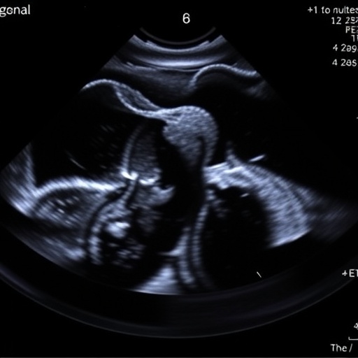
The research team addressed this gap by leveraging the enhanced temporal resolution of H-CEUS, a technique that captures extremely rapid sequences of contrast-enhanced images, enabling detailed examination of vascular patterns within lymph nodes. By intricately mapping microvascular perfusion dynamics with high frame rates, H-CEUS uncovers subtle diagnostic features undetectable with traditional imaging, offering a refined, non-invasive window into lymph node pathology.
A comprehensive patient cohort consisting of 288 individuals with enlarged superficial lymph nodes was analyzed. This group was stratified into benign (71 patients) and malignant categories (217 patients), with the malignant category further divided into metastatic (150 patients) and lymphoma subgroups (67 patients). Notably, the lymphoma cohort was subsequently classified into aggressive and indolent variants, providing insights into malignancy behavior and clinical implications. To ensure robust comparison, a subset of 67 patients from the non-lymphoma group (benign and metastatic) was randomly selected.
The researchers undertook meticulous univariate and multivariate statistical analyses to elucidate the distinguishing clinical and ultrasonographic factors associated with lymphoma. Their investigations identified several independent risk factors that strongly correlated with malignant lymphomatous involvement. Among these were advanced patient age (over 59 years), distinctive striped or reticular hyperechoic patterns detected in the lymph node cortex, and an innovative finding termed the centrifugal “fireworks” enhancement pattern observed on H-CEUS. This pattern represents a dynamic outward spread of contrast agent, reflecting aberrant vascular architecture typical of lymphoma.
In quantitative terms, H-CEUS demonstrated superior sensitivity, specificity, and overall diagnostic accuracy compared to conventional C-CEUS when cross-referenced against histopathology, the definitive diagnostic standard. The technique’s ability to discriminate between benign and malignant nodes significantly exceeded earlier modalities, illustrating the potential for H-CEUS to dramatically reduce false negatives and positives.
Beyond assessing malignancy, the study also uncovered important prognostic implications. Aggressive lymphoma subtypes exhibited markedly more heterogeneous enhancement on H-CEUS and elevated serum lactate dehydrogenase (LDH) levels, a well-known biomarker for tumor burden and cellular turnover. This correlation underscores the utility of H-CEUS not just for diagnosis but also in gauging lymphoma aggressiveness, which is pivotal for treatment planning.
The crowning achievement of this research involved the construction of a sophisticated nomogram model integrating critical clinical variables, B-mode ultrasound characteristics, and H-CEUS findings. This predictive tool succeeded in stratifying lymphoma risk with remarkable precision, evidenced by a concordance index (C-index) of 0.874. Such predictive models enhance clinical decision-making by estimating an individual patient’s likelihood of harboring lymphoma and its potential severity based on readily accessible diagnostic inputs.
Crucially, combining H-CEUS with clinical data outperformed models relying on ultrasound or clinical features alone. This synergy leverages multifaceted data points, mimicking physician diagnostic reasoning augmented by advanced imaging insights. The nomogram thus represents a scalable, user-friendly resource for clinicians aiming to optimize non-invasive assessments of superficial lymphadenopathy.
The clinical ramifications of these findings are profound. Enhanced diagnostic accuracy leads to earlier, more precise identification of lymphoma, potentially expediting biopsy decisions and appropriate oncologic referrals. By reducing diagnostic ambiguity, H-CEUS-guided workflows can minimize unnecessary invasive procedures and associated patient morbidity. Moreover, recognizing aggressive disease phenotypes aids oncologists in tailoring therapeutic intensity to individual patient risk profiles.
Despite its promise, the adoption of H-CEUS and nomogram-assisted diagnostics in routine clinical practice demands further validation across diverse populations and care settings. Integrating such advanced imaging requires investment in technology and operator expertise. However, these challenges are outweighed by the potential to shift lymphoma diagnosis paradigms toward more personalized, effective care pathways.
This study heralds a transformative era wherein dynamic imaging tools, anchored by high temporal and spatial resolution, unlock previously inaccessible details of lymph node pathology. The added layer of predictive analytics embodied in the nomogram represents a fusion of clinical acumen and data-driven innovation, embodying the future of precision oncology diagnostics.
In conclusion, the pioneering work by Song et al. demonstrates that high-frame-rate contrast-enhanced ultrasound, paired with a robust nomogram model, substantially enhances the diagnostic evaluation of superficial lymph nodes suspected of lymphoma involvement. This combined approach not only improves differentiation between benign and malignant nodes but also sheds light on lymphoma aggressiveness, paving the way for earlier intervention and tailored treatment strategies. As these technologies progress toward broader clinical implementation, they hold the promise of significantly improving patient outcomes in lymphoma care.
The scientific community awaits further exploration into the integration of H-CEUS with other emerging imaging modalities and molecular markers, potentially refining lymphoma diagnostics even further. This multidisciplinary methodology aligns seamlessly with the paradigm shift toward personalized medicine, where each patient’s unique disease characteristics inform precision therapeutics.
Ultimately, this research exemplifies how innovation in imaging technology, coupled with sophisticated analytical models, can directly impact patient care quality, illustrating a vibrant frontier in oncological diagnostics with tangible benefits for clinicians and patients alike.
Subject of Research: Diagnostic efficacy of high-frame-rate contrast-enhanced ultrasound and nomogram modeling in superficial lymphomas
Article Title: Diagnostic value of high-frame-rate contrast-enhanced ultrasound and nomogram model in lymphomas
Article References: Song, Y., Zhang, Y., Zhang, L. et al. Diagnostic value of high-frame-rate contrast-enhanced ultrasound and nomogram model in lymphomas. BMC Cancer 25, 1789 (2025). https://doi.org/10.1186/s12885-025-15074-z
Image Credits: Scienmag.com
DOI: 19 November 2025
Tags: contrast-enhanced ultrasound technologyearly detection of lymphomasenhanced temporal resolution in ultrasoundgroundbreaking cancer imaging researchhigh frame rate ultrasoundimaging techniques for lymphatic conditionslymph node characterizationlymphoma diagnosis advancementsmalignant vs benign lymph nodesnomogram model in cancer diagnosispersonalized lymphoma treatment strategiesvascular patterns in lymph nodes

