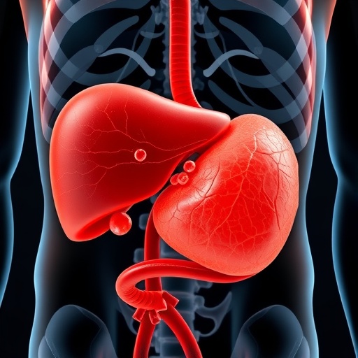
Cholelithiasis, or gallstone disease, remains a prevalent health concern across the globe, presenting a myriad of complications that can affect both adults and children. In a groundbreaking study propelled by the increasing interest in anomalies of the biliary system, researchers Sağlam and Yazıcı have unveiled a unique case involving cholelithiasis in an accessory gallbladder associated with an accessory liver. This study, published in Pediatric Radiology, opens the door to a deeper understanding of the anatomical variations that can impact gallbladder functioning, particularly in pediatric populations.
The accessory liver and its related structures are framed within the broader scope of anatomical variations that can occur in human anatomy. While many are familiar with anatomical variations such as situs inversus or the presence of additional limbs, variations in liver and biliary anatomy are less frequently acknowledged. The accessory liver, a rare phenomenon where an additional portion of liver tissue exists outside the main liver, can cause significant diagnostic challenges, especially in imaging interpretations where subtle differences can be easily overlooked.
Cholelithiasis typically occurs when there is an imbalance in the substances that make up bile, leading to the precipitation of bile salts and the formation of stones. In the context of an accessory gallbladder residing in an accessory liver, these dynamics become further complicated. The study meticulously documents the factors contributing to stone formation within this anomalous structure. The lithogenic properties of bile can be influenced by factors like bile salt concentration and gallbladder motility, significantly raising the stakes for patients with these rare anatomical configurations.
Imaging techniques have evolved exponentially to enable better identification of such rare cases. The researchers employed advanced imaging modalities such as ultrasounds, CT scans, and MRIs to conclusively identify the presence of gallstones in the accessory gallbladder. Each imaging modality brings forth its strengths; for instance, ultrasounds are often the first-line diagnostic tool due to their accessibility and non-invasive nature, while CT scans provide comprehensive structural views that highlight anomalies. The study effectively communicates the importance of utilizing a multimodal imaging approach to ensure accurate diagnosis and management of similar cases.
A major takeaway from this research is the profound implications on clinical practice. Early recognition and understanding of accessory gallbladders are crucial for the prevention of complications such as pancreatitis or biliary obstruction. Without a keen awareness of these anatomical variations, practitioners may fall into the trap of misdiagnosis, which can lead to inadequate management strategies. Proper education surrounding these rare conditions is vital for healthcare professionals to heighten their diagnostic acuity.
In light of the findings presented, the study promotes informing pediatricians and radiologists regarding the possibility of accessory gallbladders appearing in pediatric patients. Children may exhibit asymptomatic presentations, and thus, a high index of suspicion should be maintained, particularly in cases presenting with abdominal pain. The triad of gallstones, biliary colic, and anomalous anatomy may coalesce into a convoluted clinical picture that demands both patience and expertise to untangle.
The significance of genetic predisposition in gallstone formation emerges from this narrative as well. Emerging research consistently outlines a familial tendency regarding gallstone disease, yet cases like that of the accessory gallbladder challenge conventional wisdom about risk factors. Isolated anatomical variations could mean that patients classified under “low-risk” categories might still develop substantial complications. As such, genetic counseling and tailored risk assessments might become essential components in the management of affected individuals.
Equally important in this discourse is the discussion surrounding treatment options. When faced with a diagnosis of cholelithiasis in patients with accessory gallbladders, surgical intervention strategies become complex. Traditional laparoscopic cholecystectomy approaches may not apply straightforwardly, especially when the anatomy is atypical. Surgeons must prepare for uncharted territories navigated during procedures, understanding that additional structures—both anatomical and pathological—may reside beyond standard expectations.
Innovation within surgical techniques is paramount in addressing such intricate cases. Surgeons may explore minimally invasive techniques that provide greater visualization and access while minimizing trauma to surrounding tissue. Advanced robotic surgical systems may also pave the way for more sophisticated maneuvers, increasing the chances of successful outcomes while drastically reducing recovery times.
The study’s insights prompt a re-evaluation of educational curricula in medical training programs, particularly for aspiring pediatricians and radiologists. An emphasis on recognizing anatomical variations should be fundamental, yet it often remains an underrepresented component of traditional training. By furnishing future practitioners with knowledge about this field, patients could potentially avoid crises born from a lack of awareness.
Further engagement with the scientific community is encouraged to explore the connections between accessory organs and their implications for systemic health. As our understanding evolves, complementary studies that explore patient outcomes, long-term impacts, and management efficacy will be crucial. The quest for knowledge surrounding anatomical variations does not rest with singular studies; collaboration will enhance the foundational knowledge necessary to tackle these complexities adequately.
In conclusion, Sağlam and Yazıcı’s research on cholelithiasis in the accessory gallbladder of an accessory liver acts as both a call to action and a beacon of knowledge in the medical field. The intersecting realms of anatomy, diagnostics, and treatment methodologies converge to enrich our understanding of a rare pathology, revealing a landscape filled with clinical mysteries awaiting resolution.
As the scientific community continues to converge on the intersection of technology and anatomical exploration, the potential for discoveries looms large. In sporting a multidisciplinary approach, professionals across fields can break barriers built by conventional wisdom while pursuing a more holistic understanding of the human body. With studies like this, we inch towards a future where even the most elusive medical phenomena are brought to light, paving the way for more personalized and effective patient care.
Subject of Research: Cholelithiasis in the accessory gallbladder of an accessory liver.
Article Title: Cholelithiasis in the accessory gallbladder of an accessory liver.
Article References:
Sağlam, D., Yazıcı, Z. Cholelithiasis in the accessory gallbladder of an accessory liver.
Pediatr Radiol (2025). https://doi.org/10.1007/s00247-025-06440-x
Image Credits: AI Generated
DOI: https://doi.org/10.1007/s00247-025-06440-x
Keywords: Cholelithiasis, accessory gallbladder, accessory liver, anatomy, pediatric radiology.
Tags: accessory liver anomaliesanatomical variations in biliary systembiliary system health concernscholelithiasis complicationsdiagnostic challenges in imaginggallbladder functioning in childrengallstone disease prevalencegallstones in accessory gallbladderliver and biliary anatomy variationspediatric gallbladder conditionspediatric radiology studiesunique case studies in medicine


