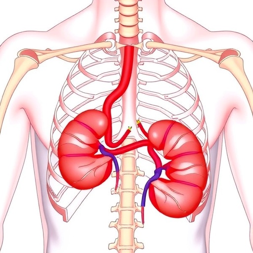
Recent advances in pediatric radiology have unveiled intricate cases that highlight the complexity of human anatomy and the unique challenges they present in medical practice. One such case involves an extremely rare condition that combines a retroaortic retrocaval reverse horseshoe kidney with distinct vertebral and spinal cord anomalies. This intriguing combination poses significant diagnostic and therapeutic challenges, as the management of each anomaly is complicated by the presence of the others. Understanding the interplay between these anatomical variations is crucial for medical professionals involved in the care of affected patients.
Horseshoe kidneys are among the most common renal fusion anomalies, characterized by the fusion of the medial aspects of the kidneys. However, the retroaortic retrocaval variant adds another layer of complexity. In typical cases, the renal vessels are situated anterior to the aorta; however, in retroaortic cases, the renal vein runs behind the aorta, which complicates surgical interventions and makes these cases particularly challenging to manage both surgically and radiologically. Thus, recognizing the retrocaval aspect on imaging becomes vital for successful intervention and treatment planning.
This retroaortic retrocaval presentation often coexists with other abnormalities, particularly in the vertebral column and spinal cord. These associated anomalies can range from minor malformations to significant developmental issues that may impact neurological function and overall health. In the described case, the presence of vertebral anomalies necessitates a comprehensive evaluation to determine their potential impact on the patient’s neurological status and how they may interact with renal function.
Imaging studies play a crucial role in the diagnosis and management of these conditions. Advanced imaging techniques such as MRI, CT, and ultrasound provide critical insights into the anatomical relationships and variations present in each patient. In this particular case, detailed imaging was paramount in elucidating the complex relationships between the renal structures and surrounding vascular anatomy. Understanding these relationships is essential for any surgical or therapeutic planning, given the high stakes of operating in such proximities.
The reviews of literature surrounding cases of retroaortic retrocaval kidneys, particularly those presenting with associated vertebral and spinal anomalies, are scant. However, the available literature indicates an increase in awareness and diagnosis of these conditions in recent years. The need for early recognition cannot be overstated; timely intervention can significantly impact patient outcomes and quality of life. The collaborative approach that includes pediatric radiologists, urologists, and orthopedic specialists offers the best chance at maximizing successful outcomes in these complex scenarios.
Despite advancements in imaging and surgical techniques, managing these congenital anomalies remains fraught with complications. Anomalies in the vertebral column can lead to varying degrees of spinal cord compression, which can manifest as symptoms ranging from pain to severe neurological deficits. Understanding the potential for these complications is vital for healthcare providers when designing a comprehensive treatment plan. The integration of multidisciplinary care is not only beneficial but often necessary to address the varied and complex needs of these patients.
In clinical practice, the implications of these findings extend to genetic counseling and long-term follow-up. The presence of renal and vertebral anomalies may indicate broader genetic syndromes that could have implications for the patient’s family. Genetic counseling can provide insights into hereditary patterns and the risk of recurrence in future pregnancies. As such, a holistic approach that considers both the immediate clinical picture and the broader biological context is invaluable.
Moreover, the interplay between the retrocaval kidney and spinal anomalies raises pertinent questions regarding embryological development. The timing and mechanisms behind renal fusion and vertebral development share intricate connections; disruptions in these processes could result in the observed anomalies. Ongoing research is necessary to glean insights into the developmental biology underlying these conditions and to elucidate potential preventative measures or therapeutic avenues.
As the medical community seeks to understand these complex cases better, continued education and research in the field of pediatric radiology are essential. Sharing unique cases within medical forums and publications can foster collaboration and enhance the collective knowledge regarding these rare presentations. It is crucial for radiologists to remain vigilant and informed about such embryological variations and their implications in pediatric populations.
In summary, the case of a retroaortic retrocaval reverse horseshoe kidney combined with vertebral and spinal cord anomalies illuminates the intricate nature of human anatomy. It challenges healthcare providers to recognize and respond to the unique needs of these patients with a comprehensive, interdisciplinary approach. This specific presentation emphasizes the importance of advanced imaging techniques and the collaborative efforts of specialized medical professionals to enhance patient outcomes.
The future of pediatric care in the context of complex anatomical variations lies in our ability to understand the deeper connections between developmental biology, clinical presentation, and management strategies. A proactive approach in education, research, and clinical practice can lead to significant advancements in diagnosing and treating such rare and multifaceted conditions.
As we look ahead, continued investigation into the genetic, environmental, and developmental factors that contribute to these anomalies will be paramount. Ensuring that final care plans are well-rounded and include considerations for the various implications of these complex conditions will ultimately pave the way for improved health outcomes for affected patients and their families.
Subject of Research: Retroaortic retrocaval reverse horseshoe kidney with associated vertebral and spinal cord anomalies.
Article Title: Retroaortic retrocaval reverse horseshoe kidney associated with vertebral and spinal cord anomalies.
Article References:
Agegn, D., Tabor, B., Buser, A. et al. Retroaortic retrocaval reverse horseshoe kidney associated with vertebral and spinal cord anomalies. Pediatr Radiol (2025). https://doi.org/10.1007/s00247-025-06399-9
Image Credits: AI Generated
DOI: https://doi.org/10.1007/s00247-025-06399-9
Keywords: Retroaortic kidney, retrocaval kidney, horseshoe kidney, vertebral anomalies, spinal cord anomalies, pediatric radiology, congenital anomalies, surgical management, imaging techniques, genetic counseling.
Tags: diagnostic imaging for renal anomalieshorseshoe kidney complicationsinterplay of anatomical variationsmanaging coexisting anatomical anomaliespediatric radiology challengesrare kidney and spine conditionsrenal fusion anomalies in childrenretroaortic retrocaval kidney anomaliesspinal cord anomalies in renal patientssurgical management of complex kidney casestherapeutic challenges in renal anomaliesvertebral column malformations

