Tumoral calcinosis is a rare condition characterized by the formation of calcium deposits in the soft tissues surrounding joint areas, particularly prevalent in individuals with underlying metabolic disorders. Understanding the complexities associated with this condition is crucial, especially when it involves critical anatomical structures like the knee joint. Recent observations have highlighted the unique epiphyseal involvement that accompanies tumoral calcinosis, underscoring its clinical significance and the need for heightened awareness among healthcare professionals.
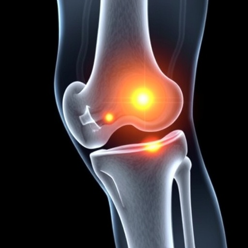
The knee joint, a vital component of human mobility, is composed of bone, cartilage, ligaments, and tendons, all of which contribute to its structural integrity and functional dynamics. In cases of tumoral calcinosis, it is essential to consider how the epiphysis—particularly the femoral and tibial epiphyses—may be affected. An understanding of the histopathological changes linked to this condition reveals that calcific masses can lead to pain, swelling, and decreased range of motion, impacting an individual’s quality of life.
Clinicians must pay close attention to the symptoms presented by patients with tumoral calcinosis. These symptoms may mimic those of more common musculoskeletal disorders, leading to diagnostic challenges. This condition often presents as palpable masses in the region of the joint, which may fluctuate in size and can be misinterpreted as other soft-tissue tumors. Awareness of the characteristic imaging features associated with tumoral calcinosis is necessary for accurate interpretation and effective management.
Radiological assessments play a pivotal role in identifying the presence of tumoral calcinosis. X-rays typically reveal well-defined, calcified lesions adjacent to the affected joint, but may not fully capture the extent of soft tissue involvement. Computed tomography (CT) and magnetic resonance imaging (MRI) provide greater detail, showcasing the soft tissue masses and their relationships with surrounding structures, which is vital for surgical planning when necessary.
Surgical intervention may be required in cases where tumoral calcinosis significantly impairs function or causes substantial discomfort. The goal of surgery is to remove the calcified masses while preserving the integrity of the surrounding tissues. Surgeons must navigate the delicate anatomy of the knee, which demands a thorough understanding of the potential risks and complications associated with such procedures.
The etiology of tumoral calcinosis is multifactorial, with detectable associations with various systemic disorders, including hyperphosphatemia and parathyroid hormone dysregulation. Further research is crucial to elucidate the underlying mechanisms driving this condition and to identify populations at higher risk. Genetic factors may also play a role, necessitating investigations into familial patterns of predisposition.
Management of tumoral calcinosis requires a multidisciplinary approach, incorporating insights from orthopedic specialists, radiologists, and metabolic disease experts. Comprehensive evaluation of the patient’s metabolic profile is essential for identifying potential underlying conditions that may contribute to the development of calcific formations. This could facilitate early intervention strategies to mitigate complications related to the condition.
Despite the rarity of tumoral calcinosis, increasing awareness and academic discourse around the condition is paramount. Continuing education for medical professionals can help prevent misdiagnoses and promote appropriate therapeutic strategies. Engaging patients in discussions about the potential need for long-term monitoring can foster a better understanding of their health management.
Current literature reflects the importance of case reports and studies to enhance the collective understanding of tumoral calcinosis, particularly in pediatric populations where growth factors may influence its manifestation and outcomes. The unique challenges presented by this condition in younger patients necessitate an informed and vigilant clinical approach.
This ongoing dialogue in the medical community about tumoral calcinosis emphasizes the importance of research, education, and awareness. As more cases are documented and studied, the complexities surrounding this condition will become clearer, enabling healthcare providers to offer improved prognoses and treatment pathways.
In conclusion, tumoral calcinosis with epiphyseal involvement in the knee represents a multifaceted clinical challenge that demands comprehensive understanding and management. Future studies are essential for illuminating the various dimensions of this condition, ultimately leading to better patient outcomes through informed medical practice and interdisciplinary collaboration.
Subject of Research: Tumoral calcinosis and its epiphyseal involvement
Article Title: Tumoral calcinosis with epiphyseal involvement in the knee
Article References:
Zhao, T., Meng, X. & Wang, Z. Tumoral calcinosis with epiphyseal involvement in the knee.
Pediatr Radiol (2025). https://doi.org/10.1007/s00247-025-06418-9
Image Credits: AI Generated
DOI: https://doi.org/10.1007/s00247-025-06418-9
Keywords: Tumoral calcinosis, epiphyseal involvement, knee, metabolic disorder, pediatric radiology, orthopedic management.
Tags: awareness among healthcare professionalscalcium deposits in soft tissuesdiagnostic challenges in musculoskeletal disordersepiphyseal involvement in tumoral calcinosishistopathological changes in calcinosisimpact on quality of life with calcinosisknee joint anatomy and functionknee tumoral calcinosismanagement of knee joint conditionsmetabolic disorders and tumoral calcinosissymptoms of tumoral calcinosistreatment options for tumoral calcinosis


