In a groundbreaking advancement for lung cancer diagnostics, researchers have developed a CT-based radiomics nomogram that incorporates adipose tissue signatures to distinguish invasive adenocarcinomas from part-solid pulmonary nodules with remarkable precision. This innovative approach represents a pivotal leap forward in the non-invasive classification of lung adenocarcinoma invasiveness, potentially transforming treatment strategies and prognostication.
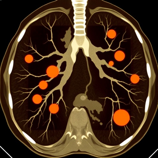
Lung adenocarcinoma remains one of the most common and deadly types of lung cancer globally, and its early and accurate assessment is critical for successful patient management. The complexity arises specifically with part-solid pulmonary nodules—lesions within the lungs that exhibit heterogeneous characteristics and are challenging to interpret using conventional imaging methodologies alone. Traditional imaging metrics provide limited information to discern invasive cancer subtypes, posing a risk of either overtreatment or undertreatment.
Recent scientific inquiry has underscored the substantial role of adipose tissue in the tumor microenvironment. Intrathoracic adipose tissue (IAT) influences various biological processes pivotal to tumor growth and invasion, including the secretion of adipokines and inflammatory mediators. Recognizing that the characteristics of adipose tissue surrounding pulmonary nodules could harbor predictive information, the research team embarked on formulating an integrative diagnostic model blending these radiomic features with established clinical parameters.
This multicenter study enlisted a cohort of 608 lung adenocarcinoma patients, collected from three distinct medical centers to ensure the method’s robustness and generalizability. High-resolution computed tomography (CT) scans were utilized to extract detailed radiomic signatures from both the pulmonary nodules and the intrathoracic adipose tissue. Employing advanced image processing algorithms, the team quantified texture, shape, intensity, and heterogeneity features that were otherwise imperceptible to the naked eye.
The core of the modelling approach was multivariable logistic regression analysis, which facilitated the development of a comprehensive nomogram—a statistical predictive tool—to classify the invasiveness of the pulmonary nodules. This nomogram included the radiomic signatures derived from the nodules and IAT, alongside key clinical factors such as nodular diameter. This multivariate approach empowered the model to capture nuanced interrelations between tumor characteristics and patient physiology.
Validation of the model’s performance was meticulous and multifaceted. The researchers established its accuracy and discriminatory capacity by analyzing the area under the receiver operating characteristic curve (AUC), a gold standard metric in diagnostic testing. Impressively, the nomogram achieved AUCs exceeding 0.9 across internal testing and two external validation cohorts, signifying excellent performance and confirming its potential applicability across diverse patient populations.
Statistical assessments including calibration metrics and Hosmer-Lemeshow goodness-of-fit tests demonstrated that the nomogram’s predictions reliably aligned with observed outcomes, confirming its clinical utility. The researchers further employed Net Reclassification Index (NRI) and Integrated Discrimination Improvement (IDI) calculations to quantify the enhancement in predictive accuracy gained when IAT radiomic features were integrated into the model. Both indices consistently indicated significant improvement, underscoring the critical value of considering adipose tissue characteristics.
Beyond raw predictive metrics, the study elevated its significance by conducting decision curve analysis, which evaluates the clinical net benefit of applying the model across a range of threshold probabilities. This analysis revealed clear advantages in guiding treatment decisions, potentially sparing patients from unnecessary surgeries or ensuring timely aggressive intervention where warranted. Moreover, stratification analyses hinted at the nomogram’s capacity to generalize beyond the derivation cohorts, offering a promising avenue for widespread clinical adoption.
This research elegantly demonstrates the power of radiomics—a field leveraging computational algorithms to extract high-dimensional quantitative features from medical images—which when synergized with biological insights about tumor microenvironments, creates precision tools tailor-made for personalized medicine. The novel incorporation of intrathoracic adipose tissue signatures marks a paradigm shift, illuminating previously overlooked tissue contexts that influence tumor behavior.
The clinical implications are profound. Accurate identification of invasive adenocarcinomas among part-solid pulmonary nodules can dramatically influence treatment pathways—dictating choices ranging from vigilant monitoring to surgical resection and adjuvant therapies. The nomogram’s ability to fine-tune risk stratification supports more nuanced, individualized patient management, potentially improving survival outcomes while mitigating unnecessary treatment risks.
Furthermore, this methodology offers a non-invasive, cost-effective alternative to invasive biopsies that carry procedural risks and sampling biases. As CT imaging is routinely performed in lung cancer screening and diagnostic workflows, the integration of radiomic analytics into existing protocols could be streamlined, facilitating rapid clinical translation without additional patient burden.
Moving forward, the research opens avenues for expanding radiomics-based models by incorporating other soft tissue types and exploring longitudinal imaging data to monitor tumor evolution. It also invites exploration of how adipose tissue signatures interact with molecular and genetic tumor profiles, potentially bridging imaging phenotypes with underlying oncogenic mechanisms.
In conclusion, this study presents a landmark advancement in lung cancer diagnostics by successfully harnessing the underappreciated radiomic signals from intrathoracic adipose tissue to enhance the differentiation of invasive adenocarcinomas within part-solid pulmonary nodules. The resulting nomogram stands as a potent, validated clinical tool that promises to inform therapeutic decision-making with unprecedented precision.
As lung cancer remains a leading cause of cancer mortality worldwide, innovations such as these are essential in the progression toward personalized oncology, where every patient’s unique disease characteristics guide treatment strategies. The integration of radiomics and adipose tissue analysis exemplifies the future of medical imaging—transforming quantitative data into actionable clinical intelligence.
Researchers anticipate that ongoing trials and real-world applications will further refine and verify the utility of this nomogram, consolidating its place in diagnostic radiology and thoracic oncology. Such cross-disciplinary collaborations between radiologists, oncologists, bioinformaticians, and data scientists continue to drive the evolution of cancer care into more predictive, preventative, and personalized paradigms.
Subject of Research:
Differentiation of invasive adenocarcinomas among part-solid pulmonary nodules using a CT-based radiomics nomogram incorporating intrathoracic adipose tissue features.
Article Title:
A CT-based radiomics nomogram incorporating adipose tissue to differentiate invasive adenocarcinomas among part-solid pulmonary nodules.
Article References:
Qin, L., Zhao, L., Li, Xm. et al. A CT-based radiomics nomogram incorporating adipose tissue to differentiate invasive adenocarcinomas among part-solid pulmonary nodules. BMC Cancer 25, 1471 (2025). https://doi.org/10.1186/s12885-025-14875-6
Image Credits: Scienmag.com
DOI:
https://doi.org/10.1186/s12885-025-14875-6
