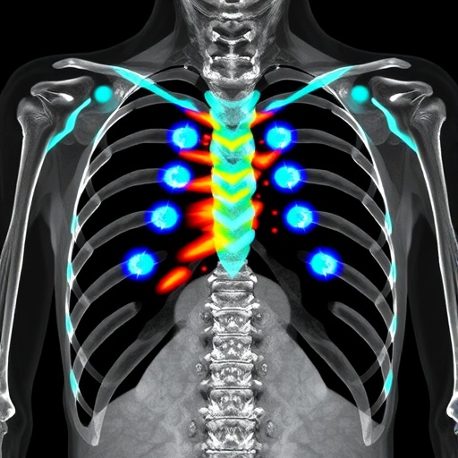
In a groundbreaking advancement poised to revolutionize forensic imaging, researchers have unveiled the remarkable diagnostic potential of post-mortem photon-counting computed tomography (PCCT) for detecting rib fractures with unprecedented accuracy. This cutting-edge technique, emerging at the intersection of forensic pathology and state-of-the-art medical imaging, offers a transformative tool for forensic investigations that demand precise skeletal assessments. Traditional imaging modalities have long struggled with subtle fracture visualization in post-mortem examinations, but PCCT ushers in a new era, promising to enhance diagnostic clarity, speed, and reliability in forensic casework.
Rib fractures, often subtle and complicated by decomposition or positioning artifacts, present a persistent challenge in medicolegal investigations. Determining the presence and pattern of such fractures can be pivotal in reconstructing injury mechanisms, clarifying circumstances surrounding death, and distinguishing between accidental trauma and foul play. Conventional computed tomography (CT) systems, while useful, frequently fall short in resolving fine osseous details post-mortem. However, PCCT leverages advanced photon-counting detectors that differ fundamentally from energy-integrating detectors used in standard CT, enabling enhanced spatial resolution and spectral imaging capabilities that prove crucial for delicate fracture detection.
At its core, PCCT represents a paradigm shift by directly counting incoming X-ray photons and categorizing them by energy level, versus traditional CT’s analog integration of all detected photons. This energy discrimination facilitates material decomposition and enhances image contrast at a microscopic scale. In the forensic context, such detailed imaging allows precise differentiation between bone, true fractures, and postmortem artifacts such as drying cracks or postmortem damage, which can otherwise mimic traumatic injury in conventional imaging. The study spearheaded by Lombardo, Hartmann, Fridle, and colleagues delves deeply into these technical advantages, offering compelling evidence of PCCT’s superior diagnostic yields.
The research undertook methodical comparisons between standard post-mortem CT scans and photon-counting CT acquisitions across a robust sample population, capturing a spectrum of trauma severities and post-mortem intervals. By meticulously correlating imaging findings with autopsy results, the investigators quantitatively analyzed sensitivity, specificity, and overall diagnostic accuracy metrics. The results unequivocally favored PCCT, which demonstrated substantially heightened sensitivity for detecting subtle cortical disruptions in ribs, many of which were missed or ambiguously interpreted on conventional CT. This validation against the autopsy gold standard underscores PCCT’s potential to refine medicolegal evidence with scientific rigor.
Beyond raw detection capabilities, PCCT also enhances image reconstruction algorithms, enabling three-dimensional volumetric analyses of fracture morphology. Such dimensions provide forensic experts with enriched contextual data, including fracture displacement, comminution, and precise anatomic localization. This comprehensive visualization enables nuanced interpretations of injury mechanisms, aiding in differentiations between blunt force trauma, postmortem artifact, and incidental skeletal conditions. The ability to integrate parametric spectral data into advanced reconstructions is an innovation unique to PCCT, offering forensic practitioners a richer diagnostic palette than ever before.
Moreover, the photon-counting mechanism inherently reduces radiation dose requirements while maintaining or improving image quality. In post-mortem imaging, where radiation exposure concerns are less restrictive than live patient scanning, this dose efficiency translates to the feasibility of high-resolution scans without prolonged acquisition times or artifact compromises. Consequently, forensic facilities can incorporate PCCT protocols into routine workflows without significant operational constraints, increasing throughput and ensuring timely medico-legal assessments critical in judicial contexts.
The study’s implications extend into forensic education and multidisciplinary collaborations. Radiologists, forensic pathologists, and imaging technologists must be trained to interpret PCCT’s nuanced outputs and spectral imaging signatures accurately. The team’s comprehensive approach includes proposed guidelines for image acquisition parameters and interpretation criteria tailored specifically for post-mortem rib assessment. Such standardization efforts are vital for harmonizing practices across forensic centers globally, facilitating consistent case comparisons and bolstering expert witness testimony reliability in legal proceedings.
Interestingly, PCCT’s advanced spectral imaging capabilities also open avenues for post-mortem tissue characterization beyond bone. While this study centers on rib fractures, the technology’s potential for soft tissue differentiation, hemorrhage visualization, and foreign body identification foreshadows a broader forensic imaging revolution. Future research may harness PCCT’s multienergy data to non-invasively explore molecular and compositional changes in tissues related to trauma, disease, or decomposition, offering deeper insights that were previously inaccessible or required invasive autopsy.
The forensic community’s response to PCCT’s emergence has been enthusiastic, recognizing its potential to overcome longstanding challenges inherent in post-mortem imaging. By bridging the gap between radiological resolution and pathological confirmation, photon-counting CT refines cause-of-death determinations and injury reconstructions with unprecedented clarity. The technique’s applicability spans homicide investigations, accident analyses, and even mass disaster victim identification, where rapid, accurate skeletal assessments can significantly influence case outcomes.
However, transitioning from pioneering research to widespread adoption entails logistical considerations. High costs of PCCT instrumentation, need for specialized software, and initial training investments must be balanced against clinical and forensic utility gains. Collaborative networks and shared facility models may help mitigate these barriers, promoting equitable access to this powerful technology. Additionally, integrating PCCT findings into multidisciplinary forensic reports requires clear communication frameworks that translate complex spectral data into actionable medico-legal conclusions comprehensible to non-specialist stakeholders, including law enforcement and juries.
In the broader scientific landscape, this breakthrough aligns with the growing trend toward precision imaging and personalized diagnostics. Just as photon-counting CT is transforming clinical radiology by improving cancer detection and cardiovascular imaging, its forensic applications underscore how technology originally conceived for medical benefit can yield profound societal impacts in justice and public safety. Lombardo and colleagues’ work exemplifies the trailblazing synergy between engineering innovation and applied forensic science, illustrating a vibrant future for imaging-enabled truth-seeking.
Ultimately, photon-counting CT’s introduction into post-mortem forensic practice heralds an era where accurate, non-destructive rib fracture detection and skeletal trauma mapping become routine rather than exceptional. This leap forward promises to elevate autopsy standards, reduce diagnostic uncertainty, and strengthen the evidentiary foundation of medico-legal investigations worldwide. The ripple effects extend beyond technical improvements; they pave the way for ethical, transparent, and scientifically robust death investigations that uphold justice and honor the dignity of the deceased.
As forensic imaging continues to evolve, the integration of photon-counting CT will likely inspire further technological innovations, including artificial intelligence-assisted image interpretation, automated fracture detection algorithms, and multi-modal forensic datasets that fuse anatomical, biochemical, and spectral information. Each of these advancements, rooted in the diagnostic breakthroughs demonstrated by Lombardo, Hartmann, Fridle, and their team, brings the forensic sciences closer to a future where truth is uncovered with unparalleled precision, speed, and confidence.
The study’s findings ripple through not only forensic medicine but also forensic anthropology, radiology, and pathology disciplines, emphasizing multidisciplinary collaboration as critical to fully harnessing photon-counting CT’s promise. As institutions worldwide consider the next generation of forensic imaging workflows, this technology’s robust performance in rib fracture detection underscores its role as a cornerstone innovation, redefining what is possible at the intersection of technology and justice.
In conclusion, the application of photon-counting computed tomography to forensic post-mortem rib fracture detection marks a pivotal advancement in forensic imaging science. Lombardo et al.’s rigorous research delineates a clear pathway from innovative imaging physics to impactful forensic practice. This new modality offers a potent combination of enhanced diagnostic accuracy, improved artifact differentiation, dose efficiency, and rich spectral data that collectively transform the landscape of post-mortem examinations. Its adoption promises to enhance the evidentiary value of forensic investigations, safeguard judicial processes, and ultimately contribute to the pursuit of truth through science.
Article Title:
Diagnostic accuracy of rib fracture detection in forensic post-mortem photon counting CT
Article References:
Lombardo, P., Hartmann, C., Fridle, C. et al. Diagnostic accuracy of rib fracture detection in forensic post-mortem photon counting CT. Int J Legal Med (2025). https://doi.org/10.1007/s00414-025-03597-w
Image Credits: AI Generated
Tags: challenges in medicolegal imagingdiagnostic clarity in forensic investigationsdistinguishing accidental trauma from foul playenergy-integrating vs photon-counting detectorsenhancing forensic casework accuracyforensic pathology advancementsphoton-counting computed tomographypost-mortem imagingreconstructing injury mechanismsrib fracture detectionskeletal assessments in forensicssubtle fracture visualization techniques


