In recent developments in the field of imaging technology, a novel method using multi-energy X-ray imaging has emerged, showcasing its potential for various applications. This innovative technique allows for the distinction of materials based on their composition and density, thus enhancing the capabilities of traditional imaging methods. The implications of this technology stretch across several key domains, including medical diagnostics and material analysis, emphasizing the importance of the advancements being made in this arena.
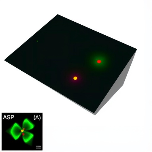
The crux of this advancement lies in the development of polymer-ceramic composite scintillator films. These films possess specific responses to different energy ranges of X-rays, effectively enabling the imaging technology to discern various objects based on their material properties. The unique combination of materials used in these films contributes not only to their performance but also to the efficiency and accuracy of the imaging process. The integration of a vitamin-assisted in-situ growth method further enhances these scintillator films’ quality, as demonstrated by the research led by Prof. Menglu Chen from the Beijing Institute of Technology.
The research group’s innovative approach incorporates vitamin B1 (VmB1) in the synthetic process of the scintillator films. This method promotes high uniformity and radiation stability over extended periods, essential characteristics for any reliable imaging technology. The in-situ growth method facilitates the formation of perovskite polymer-ceramic films that maintain the desired properties necessary for effective X-ray imaging. This technique also simplifies the material selection process, traditionally a significant obstacle in the development of multi-energy X-ray imaging systems.
Calculations and experimental data support the efficacy of the proposed method. The research team deployed density functional theory (DFT) to evaluate the charge distribution among polymer functional groups in conjunction with perovskite materials. Findings suggest a substantial relationship between the interaction energy of polyvinyl alcohol (PVA) and the perovskite, which directly enhances the optical properties of the resulting scintillator films. By leveraging this knowledge, the team has laid out a framework for selecting polymer hosts, thus facilitating future research and application of similar technologies.
Furthermore, the architecture of the scintillator films plays a crucial role in the efficiency of multi-energy X-ray imaging. The research highlights a systematic approach to designing the type, thickness, and stacking sequence of the layers comprising the scintillator films. Such meticulous planning ensures that the films are capable of capturing accurate data across varying energy levels, crucial for a reliable multi-energy imaging system. Different absorption distributions across the layers of the films were meticulously calculated, bringing clarity to the design specifications needed to attain optimal imaging resolution.
The culmination of this research is the successful development of a four-channel multi-energy X-ray imaging system. The apparatus operates over an energy range of 10 keV to 60 keV, representing a significant achievement in the field of imaging technology. The ability to distinguish between materials such as metal and plastic with clarity from a single X-ray shot demonstrates the system’s robustness and versatility in practical applications. Additionally, the use of flexible polymer-ceramic scintillator films allows for high-resolution imaging of curved objects, a feat not easily achieved with traditional imaging systems.
The introduction of this technology brings substantial benefits to industries requiring advanced imaging capabilities. For instance, in the medical field, this method can enhance diagnostic accuracy by providing detailed insights into biological structures, enabling better detection of anomalies. Moreover, the capacity to differentiate materials based on subtle density differences expands the potential for innovation in various engineering and industrial sectors, where precision material handling and analysis is critical.
As more researchers and practitioners recognize the significance of multi-energy X-ray imaging, it is likely that we will see a proliferation of its applications. The ability to visualize and differentiate materials with unparalleled clarity opens new avenues for research and development. By continuing to refine and expand upon these initial findings, the scientific community can push the boundaries of imaging technologies further, paving the way for discoveries that were previously considered unattainable.
In light of these developments, a publication in the esteemed journal PhotoniX has detailed this research, sparking interest across multiple scientific disciplines. The article provides insights into the methodologies employed, the results obtained, and the potential implications for future work. Researchers, industry practitioners, and scholars alike will benefit from the shared knowledge and proposed advancements in this field, as it underscores the importance of collaborative efforts in fostering innovation.
The pursuit of advancing imaging technologies, bolstered by rigorous research and innovative methodologies, highlights the ongoing quest for improved understanding and manipulation of material properties. Through the successful implementation of techniques like the vitamin-assisted in-situ growth method, researchers are setting the stage for a new era of imaging technology, with the potential to redefine how we perceive and analyze the world around us.
The multi-energy X-ray imaging system represents not only a significant technological breakthrough but also serves as a foundation for future exploration in related fields. As attention shifts toward enhancing imaging capabilities, the emphasis on interdisciplinary research will be crucial in finding solutions to longstanding challenges. With continued investment and curiosity, the scientific community is poised to uncover new insights, making this an exciting period of advancement in imaging technology.
This groundbreaking work stands as a testament to the power of innovation and collaboration in scientific research. By integrating novel approaches with established techniques, researchers are carving out new pathways for understanding complex material interactions. As the field progresses, the anticipation for further developments in multi-energy X-ray imaging continues to grow, promising an array of applications that could revolutionize various sectors.
Subject of Research: N/A
Article Title: In-situ grown polymer-ceramic scintillator and applications on X-ray multi-energy curved surface imaging.
News Publication Date: 12-Aug-2025
Web References: N/A
References: N/A
Image Credits: Menglu Chen
Keywords
Multi-energy X-ray imaging, polymer-ceramic scintillator films, vitamin-assisted growth method, imaging technology, density functional theory, innovative research, optical properties, imaging resolution, medical diagnostics, material analysis, scientific collaboration.
Tags: advanced multi-energy X-ray imagingcomposite materials in X-ray applicationsenergy range discrimination in imagingImaging technology advancementsin-situ grown multi-layer scintillatorsmaterial analysis techniquesmedical diagnostics imaging technologypolymer-ceramic composite scintillator filmsProf. Menglu Chen research contributionsradiation stability in imagingscintillator film performance enhancementvitamin-assisted growth methods


