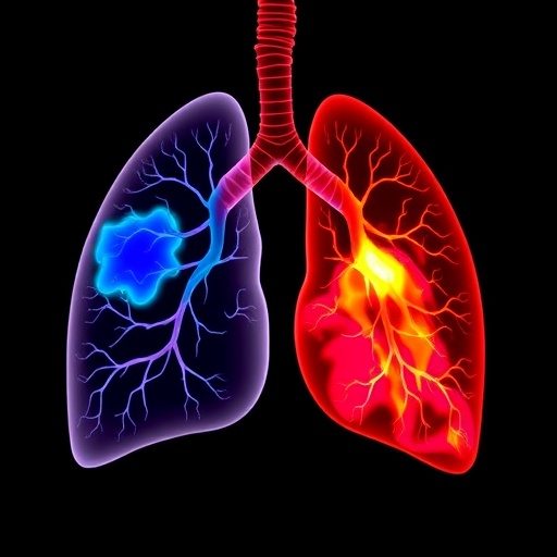

In the realm of medical imaging and oncology, the precise delineation of tumor boundaries in lung cancer remains a critical, yet formidable challenge. A recent breakthrough, documented in the journal BMC Cancer, introduces a pioneering deep learning framework known as D-S-Net that promises to significantly enhance the accuracy and efficiency of Gross Tumor Volume (GTV) segmentation in lung cancer CT scans. This innovation taps into the expanding potential of artificial intelligence in clinical diagnostics and treatment planning, particularly in radiotherapy where precise tumor mapping is paramount.
Lung cancer, notorious for its complexity due to the heterogeneity of tumor morphology and often subtle boundaries in CT imaging, poses substantial obstacles for effective segmentation algorithms. The high-resolution CT images provide detailed anatomical information but require sophisticated processing to distinguish tumor from normal tissue. Historically, segmentation has been hampered by the small size of tumors and ambiguous edges, which reduce the reliability of automated methods and force reliance on time-consuming manual delineations by expert radiologists.
To address these challenges, the architects of D-S-Net conceived a dual-stage network strategy that leverages a division of labor between tumor detection and fine-grained segmentation. The first phase is dedicated to swiftly pinpointing candidate regions within high-resolution 512×512 pixel CT slices using a streamlined detection network. This step tactically narrows down the data fed into the subsequent segmentation stage, substantially lowering computational overhead without sacrificing input resolution or detail, a common pitfall in many existing models.
Following this, the second phase of D-S-Net applies a modified U-Net architecture, a widely recognized convolutional neural network framework known for image segmentation tasks, customized here to operate on the localized tumor regions identified earlier. This stage incorporates a spatial attention mechanism, which dynamically focuses the model’s “awareness” on relevant spatial features, thereby refining segmentation precision by enhancing the contrast between tumor and surrounding tissues.
An integral component of the D-S-Net design is its innovative loss function scheme, which combines binary cross-entropy and Dice loss metrics. This hybrid loss effectively counters the class imbalance problem that pervades medical image segmentation, where the disproportion between tumor pixels and the vast background often misguides naive optimization approaches. By uniting these two metrics, the network is encouraged to balance pixel-wise accuracy with region overlap fidelity in its segmentation output.
The model’s evaluation on a comprehensive lung cancer GTV dataset revealed a significant leap in segmentation performance. D-S-Net achieved a Dice coefficient of 78.52%, marking a 5.49% improvement over the previously best-performing SwinU-Net model. This metric quantitatively reflects the overlap between predicted tumor regions and ground truth annotations, underscoring the model’s enhanced delineation capability. Furthermore, when tested on a second independent dataset, the model’s performance soared even higher, obtaining a Dice coefficient of 86.56%—outstripping its competitor by a remarkable 13.19%.
Beyond mere accuracy, extensive ablation studies elucidated the critical influence of each architectural choice on the final results. The detection stage not only improves efficiency by focusing computational resources but also enhances overall accuracy by filtering irrelevant areas. The spatial attention mechanism proved indispensable for discriminating fine tumor boundaries, and the synergistic loss function was pivotal in maintaining balance between sensitivity and specificity in voxel classification. Collectively, these components form a synergy that elevates both the reliability and practicality of the segmentation process.
Importantly, the computational complexity analysis presented in the study confirmed that D-S-Net is not only precise but also efficient. In a clinical context, where fast image processing is essential for real-time decision-making and adaptive radiotherapy, the model balances speed and performance adeptly. This efficiency stems from the dual-stage approach, where heavy computation is reserved solely for promising regions, a strategy that also facilitates potential scalability and integration with existing hospital imaging workflows.
The clinical implications of this work are profound. Accurate GTV segmentation underpins optimal radiation dosage planning, directly impacting treatment efficacy and minimizing collateral damage to healthy lung tissue. The introduction of D-S-Net could alleviate radiologists’ workload and reduce inter-observer variability, a significant source of inconsistency in treatment outcomes. Moreover, its adaptability suggests potential applications beyond lung cancer, in other oncological settings where tumor precision mapping is equally vital.
The methodology employed by the research team demonstrates a sophisticated understanding of the interplay between detection and segmentation within a deep learning context. By treating localization and boundary refinement as complementary tasks, D-S-Net mimics the diagnostic reasoning of expert clinicians who first identify suspicious regions before scrutinizing them in detail. This intelligent design philosophy marks a paradigm shift from monolithic network approaches that attempt to tackle the entire segmentation problem in one step.
Furthermore, the incorporation of spatial attention mechanisms reveals how insights from cognitive neuroscience and computer vision can be effectively harnessed in medical imaging. By enabling dynamic prioritization of spatial features, the network better captures subtle variations in tumor appearance, which are critical in distinguishing malignant growths from surrounding tissues like fibrosis or atelectasis that often confound less specialized algorithms.
The hybrid loss function methodology also deserves particular emphasis. The combined binary cross-entropy and Dice loss addresses the common issue of imbalanced datasets in medical imaging, where background voxels vastly outnumber tumor voxels. This balanced optimization ensures that the network does not simply favor the majority class but learns a nuanced representation that recognizes the spatial extent of tumors with higher sensitivity, thus avoiding undersegmentation.
While this study represents a significant advancement, the authors acknowledge avenues for further refinement. Incorporation of multi-modal imaging data such as PET-CT or MRI could enhance tumor characterization further, while adaptation to 3D volumes might leverage spatial context more comprehensively. Additionally, prospective clinical trials are necessary to validate the real-world impact of D-S-Net on patient outcomes and workflow integration.
In conclusion, the D-S-Net framework embodies a strategic fusion of modern deep learning architecture, computational efficiency, and clinical relevance. It stands as an exemplar of how targeted network design paired with domain-specific innovations like spatial attention and combined loss functions can produce tangible benefits in the demanding field of medical image analysis. As lung cancer continues to challenge clinicians worldwide, such AI-driven tools offer a promising horizon for improving treatment precision and patient quality of life.
Subject of Research: High-precision segmentation of Gross Tumor Volumes (GTV) in lung cancer CT images using deep learning.
Article Title: D-S-Net: an efficient dual-stage strategy for high-precision segmentation of gross tumor volumes in lung cancer CT images
Article References:
Yi, C., Jiang, S., Xiong, L. et al. D-S-Net: an efficient dual-stage strategy for high-precision segmentation of gross tumor volumes in lung cancer CT images. BMC Cancer 25, 1387 (2025). https://doi.org/10.1186/s12885-025-14615-w
Image Credits: Scienmag.com
DOI: https://doi.org/10.1186/s12885-025-14615-w
Tags: artificial intelligence in oncologyautomated tumor detection algorithmschallenges in lung cancer imagingD-S-Net lung tumor segmentationdeep learning in medical imagingdual-stage neural network for segmentationGross Tumor Volume segmentationhigh-resolution CT scan analysisimproving accuracy in cancer diagnosticsprecise tumor boundary delineationradiotherapy tumor mappingsegmentation of heterogeneous tumors
