
In recent years, the quest for more accurate and less invasive diagnostic tools in oncology has accelerated, particularly in the field of dermatology where skin cancer remains the most prevalent malignancy worldwide. Conventional diagnostic methods often depend heavily on visual inspection followed by invasive biopsies, which, while effective, can be time-consuming, uncomfortable, and carry the risk of scarring or infection. Addressing these limitations, pioneering research spearheaded by teams at Saint-Étienne University Hospital and Paris-Saclay University in collaboration with Damae Medical in France has culminated in a breakthrough: a compact, noninvasive imaging system that synergistically integrates line-field confocal optical coherence tomography (LC-OCT) and confocal Raman microspectroscopy. This dual-modality apparatus ushers in a new era of skin cancer detection by concurrently unraveling the morphological and chemical nuances of cancerous tissues at the cellular level.
At the technological core, LC-OCT offers unprecedented high-resolution images of skin microstructures, capturing cellular architecture in vivo with exquisite detail. Unlike traditional optical coherence tomography systems, LC-OCT operates through a line-field scanning approach that enhances image speed and depth penetration, allowing clinicians to visualize hierarchical tissue structures spanning from the stratum corneum down to the dermis. This meticulous structural mapping facilitates the localization of potentially malignant lesions with remarkable precision. However, morphological examination alone can be insufficient to demarcate cancerous from benign tissues in ambiguous cases, necessitating complementary analytical methods.
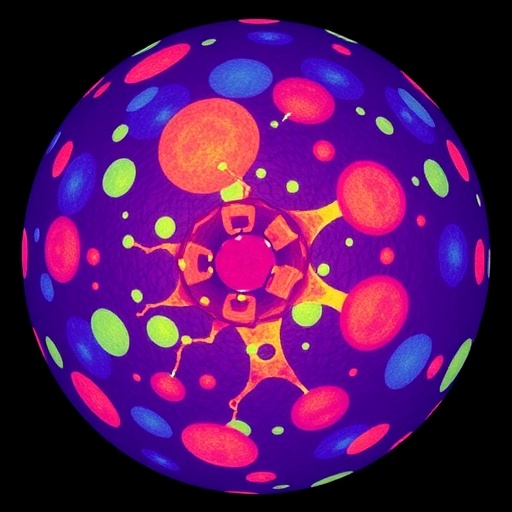
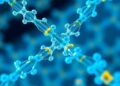
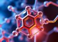
To bridge this gap, the research team incorporated confocal Raman microspectroscopy into the imaging platform. Raman spectroscopy capitalizes on inelastic scattering of monochromatic light to yield molecular fingerprints of tissues. By focusing laser light on microscopic regions flagged by LC-OCT, this technique elucidates the chemical compositions underlying morphological abnormalities. The resulting Raman spectra reveal subtle variations in biomolecular constituents such as lipids, proteins, and nucleic acids, which can differ markedly between healthy and neoplastic cells. Integrating these chemical signatures with high-resolution structural data significantly enriches the diagnostic landscape.
.adsslot_LCyTF3M5tP{width:728px !important;height:90px !important;}
@media(max-width:1199px){ .adsslot_LCyTF3M5tP{width:468px !important;height:60px !important;}
}
@media(max-width:767px){ .adsslot_LCyTF3M5tP{width:320px !important;height:50px !important;}
}
ADVERTISEMENT
Transitioning from concept to clinic, the system underwent rigorous validation over the course of one year involving more than 330 skin cancer samples, predominantly encompassing nonmelanoma variants like basal cell carcinoma (BCC) and squamous cell carcinoma (SCC). Initially, LC-OCT was employed to pinpoint suspicious microstructural formations within the tissue. Subsequent application of confocal Raman microspectroscopy generated an extensive dataset comprising over 1,300 chemical spectra sampled from these delineated areas. These spectra represent a complex multidimensional dataset requiring sophisticated analytical techniques to interpret.
To manage and decode this intricate information, the investigators harnessed artificial intelligence, training machine learning algorithms to recognize patterns indicative of cancerous versus normal tissues. The AI model was calibrated on annotated spectral and imaging datasets, enabling it to autonomously classify the lesions. Impressively, the model achieved an accuracy rate of 95% in identifying basal cell carcinoma and maintained a robust 92% accuracy when encompassing both BCC and SCC classifications. Such performance underscores the promise of AI-augmented imaging in dermatological diagnostics.
Beyond diagnostic precision, the dual-imaging approach unveiled fundamental insights into the biochemical diversity among skin cancer subtypes. Distinct chemical fingerprints linked to different carcinomas emerged from spectral analyses, shedding light on the molecular underpinnings of tumor development and progression. This deeper understanding opens avenues for personalized treatment regimens tailored to the biochemical idiosyncrasies of each cancer type, potentially enhancing therapeutic efficacy.
One of the most compelling attributes of this system is its noninvasive nature. Patients benefit from a painless procedure that obviates the need for surgical biopsy in many cases, reducing physical and psychological burdens. Moreover, the rapid turnaround time for image and spectral acquisition could enable clinicians to make more immediate diagnostic and treatment decisions, effectively streamlining patient management workflows.
From a clinical perspective, the amalgamation of structural and chemical insights obtained by this innovative instrument represents a paradigm shift. Whereas histopathology remains the gold standard, it requires tissue extraction and processing that impose inevitable delays. In contrast, this dual-modality technique furnishes near real-time evaluations at the bedside, facilitating earlier interventions. Furthermore, the system’s compactness ensures compatibility with standard dermatology practices without necessitating cumbersome or costly infrastructure upgrades.
The implications of these findings resonate beyond dermatology, signaling a broader trend toward convergence of multimodal imaging and AI in medical diagnostics. The seamless integration of nanoscale structural imaging with molecular spectroscopy, empowered by artificial intelligence, exemplifies the future trajectory of precision medicine. By corroborating morphological anomalies with biochemical context, clinicians are equipped with richer datasets from which to derive diagnoses with heightened confidence.
Looking ahead, the research group envisions further refinements to enhance spectral resolution and imaging depth, as well as expansion of the AI training library with more diverse data to bolster generalizability across populations and cancer subtypes. Scaling these innovations to widespread clinical adoption will necessitate collaborative efforts among engineers, clinicians, and regulatory bodies, but the foundation laid by this study is robust and promising.
Ultimately, the combination of LC-OCT and confocal Raman microspectroscopy heralds a new frontier in skin cancer diagnostics. This integrative approach not only improves diagnostic accuracy but also ushers in minimally invasive protocols that prioritize patient comfort without compromising clinical outcomes. As skin cancer incidence continues to rise globally, innovations such as these are vital to enhancing early detection, refining diagnostic precision, and ultimately reducing mortality.
The study, slated for publication in the Journal of Biomedical Optics, stands as a testament to multidisciplinary collaboration and technological ingenuity in healthcare. It exemplifies how cutting-edge physics and chemistry can be translated into tools that tangibly improve patient care. With ongoing advancements, the dream of real-time, noninvasive, and highly accurate cancer diagnostics moves closer to becoming a clinical reality.
Subject of Research: Nonmelanoma skin cancer identification using combined imaging and spectroscopy techniques.
Article Title: AI-assisted identification of nonmelanoma skin cancer structures based on combined line-field confocal optical coherence tomography and confocal Raman microspectroscopy
News Publication Date: 28-Jul-2025
Web References:
https://www.spiedigitallibrary.org/journals/journal-of-biomedical-optics/volume-30/issue-07/076008/AI-assisted-identification-of-nonmelanoma-skin-cancer-structures-based-on/10.1117/1.JBO.30.7.076008.full
References:
M. Ayadh et al., “AI-assisted identification of nonmelanoma skin cancer structures based on combined line-field confocal optical coherence tomography and confocal Raman microspectroscopy,” J. Biomed. Opt. 30(7), 076008 (2025), doi:10.1117/1.JBO.30.7.076008.
Image Credits: M. Ayadh et al., doi 10.1117/1.JBO.30.7.076008
Keywords
Imaging, High resolution imaging, Skin cancer, Diagnostic imaging, Optical coherence tomography, Raman spectroscopy, Chemical analysis
Tags: cellular-level cancer analysiscompact imaging system for dermatologyconfocal Raman microspectroscopydermatology diagnostic toolsdual-mode optical imaging systemhigh-resolution skin imaginginnovative oncology researchline-field confocal optical coherence tomographynoninvasive skin cancer diagnosisParis-Saclay University collaborationSaint-Étienne University Hospitalskin cancer detection technology

