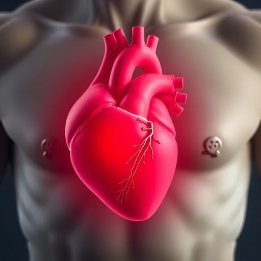
Epicardial adipose tissue (EAT) has long been relegated to the sidelines of cardiovascular research, considered merely a passive fat depot nestled around the heart. However, emerging evidence is rapidly transforming our understanding of this tissue from a biological bystander into a dynamic and multifaceted organ that exerts profound influences on cardiac function and pathology. This tissue, sandwiched between the myocardium and visceral pericardium, wields an astonishing range of biochemical and mechanical effects, oscillating between protective and harmful roles. The recent comprehensive review by Tian, Wang, and Dong elucidates these paradoxical functions of EAT, charting a nuanced course through its complex biology and divergent impacts on cardiovascular health.
At face value, epicardial adipose tissue acts as a metabolic powerhouse directly supplying the myocardium with essential fatty acids and energy substrates, facilitating efficient cardiac performance under physiological conditions. Far from being a mere fat store, EAT exhibits remarkable plasticity, producing anti-inflammatory adipokines and bioactive molecules that temper local immune responses. This immunomodulatory capacity positions EAT as a frontline sentinel safeguarding myocardial integrity, cushioning the heart mechanically and buffering coronary vessels from exaggerated inflammatory and oxidative insults. These protective features underscore a sophisticated crosstalk between EAT and cardiac cells that promotes homeostasis and resistance to stress.
Yet this harmony is exceedingly delicate. Pathological perturbations, such as obesity, diabetes, or chronic inflammation, can induce a pernicious transformation within EAT’s cellular milieu. Instead of secreting salutary factors, EAT turns into an insidious perpetrator, releasing potent pro-inflammatory cytokines, chemokines, and reactive oxygen species (ROS) that propagate oxidative stress and endothelial dysfunction. These noxious signals permeate through the thin epicardial layer, directly impacting adjacent myocardium and coronary arteries. This biochemical assault is now recognized as a critical driver fueling the progression of atherosclerosis, coronary artery disease, and arrhythmogenesis, including atrial fibrillation. In essence, EAT embodies a double-edged sword in cardiovascular disease pathophysiology.
.adsslot_ul5gknLKNE{width:728px !important;height:90px !important;}
@media(max-width:1199px){ .adsslot_ul5gknLKNE{width:468px !important;height:60px !important;}
}
@media(max-width:767px){ .adsslot_ul5gknLKNE{width:320px !important;height:50px !important;}
}
ADVERTISEMENT
Digging deeper into the molecular mechanisms, research highlights that EAT harbors an intricate cellular heterogeneity comprising adipocytes, immune cells (macrophages, T-cells), fibroblasts, and endothelial elements. This complex cellular ensemble orchestrates a delicate balance of pro- and anti-inflammatory signals depending on physiological context. The shift toward a pro-inflammatory phenotype is marked by infiltration of M1-like macrophages that secrete tumor necrosis factor-alpha (TNF-α), interleukin-6 (IL-6), and other deleterious mediators. These factors not only exacerbate local inflammation but also induce fibroblast activation and myocardial fibrosis — hallmarks of adverse cardiac remodeling and heart failure. Understanding how these cells communicate and transform EAT’s behavior offers fertile ground for therapeutic innovation.
One particularly captivating focus involves EAT’s metabolic flexibility and thermogenic potential. Contrary to white adipose depots, EAT exhibits features akin to beige or brown fat, capable of dissipating energy through uncoupling protein 1 (UCP1)-mediated thermogenesis. This capacity for adaptive heat generation and substrate oxidation may shield the heart from lipotoxicity and metabolic derangements. However, under disease conditions, these beneficial functions wane, leading to lipid overload and mitochondrial dysfunction within EAT. Such metabolic reprogramming not only impairs energy provision but also fuels local inflammatory cascades. Deciphering these metabolic switchpoints is paramount to harnessing EAT’s protective functions.
Importantly, the anatomical proximity of EAT to vital coronary vessels enables its paracrine and vasocrine signaling to directly affect vascular tone and remodeling. Pro-inflammatory cytokines and adipokines from EAT can penetrate the vasa vasorum and adventitia of coronary arteries, promoting endothelial activation, smooth muscle proliferation, and plaque instability. This unique spatial arrangement blurs traditional compartmental boundaries and underscores EAT’s active role in sculpting coronary atherosclerotic lesions. Advanced imaging techniques now allow quantification and characterization of EAT volume and activity, revealing strong correlations with coronary artery disease severity and prognosis, thereby cementing its clinical relevance.
Atrial fibrillation (AF), the most common sustained cardiac arrhythmia, also finds etiological roots in pathological modifications of EAT. Increased EAT volume and inflammation correlate with atrial structural remodeling, fibrosis, and electrical conduction abnormalities. The infiltration of immune cells and pro-fibrotic mediators from EAT onto the atrial myocardium enhances arrhythmogenic substrate formation. This mechanistic link opens exciting potential for targeted interventions aiming to modulate epicardial fat inflammation and reduce arrhythmia burden, potentially transforming clinical management paradigms for AF.
Modern technological advances have accelerated research unraveling EAT’s biology, employing multi-omics analyses, single-cell sequencing, and novel in vivo imaging modalities. These tools reveal previously unappreciated heterogeneity within EAT, with distinct subpopulations exhibiting divergent functional signatures. Such discovery paves the way for precision medicine approaches that could selectively reprogram EAT’s secretory profile, favoring cardioprotective over deleterious outputs. The therapeutic landscape may eventually encompass anti-inflammatory agents, metabolic modulators, and gene therapies specifically designed to recalibrate EAT’s influence on cardiac and vascular tissues.
Despite remarkable progress, significant knowledge gaps remain. The triggers instigating EAT’s phenotypic switch, the temporal sequence of molecular events, and the interplay with systemic metabolic status are not fully elucidated. Moreover, inter-individual variability influenced by genetics, sex differences, and environmental factors complicate the translation of findings into broad clinical applications. Longitudinal studies and well-controlled interventions are needed to untangle causal relationships and validate biomarkers that reliably reflect EAT-related cardiovascular risk.
Furthermore, the potential for therapeutic targeting remains tantalizing yet challenging. Global adiposity reduction strategies via lifestyle or pharmacologic interventions may partially mitigate EAT-associated risks, but more precise approaches are required to avoid unintended consequences. Innovations such as localized drug delivery, immune modulation, or bioengineered scaffolds offer futuristic avenues to remodel EAT’s cellular microenvironment. A comprehensive understanding of the molecular cross-talk between EAT and cardiac tissues is pivotal to overcome these translational hurdles.
In clinical contexts, the measurement of EAT volume and activity is gaining traction as a non-invasive biomarker to stratify cardiovascular risk. Techniques such as cardiac magnetic resonance imaging (MRI), computed tomography (CT), and echocardiography are increasingly employed to assess EAT’s contribution to coronary disease and arrhythmias. Incorporating EAT metrics into existing risk assessment models may enhance predictive accuracy and enable personalized therapeutic strategies. This paradigm shift heralds a novel dimension of cardiovascular diagnosis and prognosis.
In conclusion, epicardial adipose tissue emerges not as a mere anatomical curiosity but as a central player in cardiovascular health and disease. Its Janus-faced nature—where a guardian role can swiftly transform into a harmer—reflects the exquisite cellular and molecular complexity modulated by systemic and local context. By illuminating these dualistic mechanisms, the recent review galvanizes scientific and clinical communities to reimagine the heart’s surrounding fat as a therapeutic target. Ultimately, unlocking the secrets of EAT promises to revolutionize cardiovascular medicine, presenting both challenges and unprecedented opportunities in combating heart disease.
As the field evolves, interdisciplinary collaboration between cardiologists, immunologists, metabolic researchers, and bioengineers will be vital to translate bench discoveries into tangible therapies. The epicardial adipose tissue—the heart’s enigmatic neighbor—stands as a frontier in cardiovascular research, reminding us that sometimes the closest tissues hold the most profound secrets. Harnessing its protective capacity while mitigating its harmful tendencies may pave the way to healthier hearts worldwide.
Subject of Research: Epicardial adipose tissue and its dual role in cardiovascular health and disease.
Article Title: Heart-guarding or heart-harming? The dual role of epicardial adipose tissue in cardiovascular health and disease.
Article References:
Tian, Y., Wang, P. & Dong, Z. Heart-guarding or heart-harming? The dual role of epicardial adipose tissue in cardiovascular health and disease. Int J Obes (2025). https://doi.org/10.1038/s41366-025-01852-z
Image Credits: AI Generated
DOI: https://doi.org/10.1038/s41366-025-01852-z
Tags: adipokines and cardiovascular functioncardiovascular research on epicardial fatcrosstalk between epicardial fat and myocardiumdynamics of epicardial fatepicardial adipose tissue functionsheart health and epicardial fatimmunomodulation by epicardial adipose tissueinflammatory responses in heart healthmechanical cushioning of the heartmetabolic roles of epicardial fatprotective effects of epicardial fatrole of epicardial fat in heart disease


