A groundbreaking luminescent sensor designed for detecting the liver cancer biomarker β-glucuronidase has emerged from the laboratories of the Indian Institute of Science (IISc). This innovative system utilizes a terbium-based paper sensor that offers an unprecedented combination of sensitivity, simplicity, and accessibility. The research team’s novel approach not only promises to transform early cancer diagnostics but also brings hope for cost-effective screening in resource-limited settings worldwide. Leveraging the unique photophysical properties of terbium, a rare earth metal, this sensor targets β-glucuronidase, an enzyme intricately linked to multiple pathological conditions, including liver cancer.
β-glucuronidase is an evolutionarily conserved enzyme, omnipresent in a diverse array of life forms from microbes to mammals. Biochemically, its primary function involves hydrolyzing glucuronic acid conjugates, acting on glycosidic bonds within various substrates. Clinically, its elevated expression serves as a biomarker indicating the presence of several diseases. Notably, increased β-glucuronidase levels correlate with liver cancer progression, as well as other malignancies such as colon, breast, and renal cancers. The enzyme’s activity is also heightened in infectious states like urinary tract infections and immunodeficiency syndromes including AIDS, reinforcing its medical significance as a diagnostic target.
Traditional enzyme detection methodologies, such as colorimetric assays and conventional fluorescence techniques, have long been hindered by limitations in sensitivity and susceptibility to background interference. Fluorescent probes typically suffer from short-lived excited states, which overlap with endogenous autofluorescence, complicating signal interpretation. The research team ingeniously circumvents these issues by employing terbium ions, known for their exceptionally long-lived luminescent excited states. This unique trait enables temporal separation of the desired luminescent signal from the background noise, vastly improving detection clarity and accuracy.
.adsslot_U6VJhvo90j{ width:728px !important; height:90px !important; }
@media (max-width:1199px) { .adsslot_U6VJhvo90j{ width:468px !important; height:60px !important; } }
@media (max-width:767px) { .adsslot_U6VJhvo90j{ width:320px !important; height:50px !important; } }
ADVERTISEMENT
The conceptual foundation of this sensor lies in the chemistry of rare earth metal luminescence combined with targeted enzymatic activation. Terbium ions are embedded within a specially formulated gel matrix derived from bile salts, creating a stable fluorescent environment. The gel serves both as a scaffold to hold terbium ions in proximity and as a medium facilitating efficient energy transfer processes. An organic molecule, 2,3-Dihydroxynaphthalene (2,3-DHN), chemically masked with glucuronic acid, is incorporated into this matrix. When β-glucuronidase enzymatically cleaves the glucuronic acid moiety, free 2,3-DHN is released and acts as an antenna to sensitize terbium luminescence.
The operational mechanism is elegantly straightforward yet sophisticated. Upon UV light excitation, free 2,3-DHN absorbs energy and transfers it efficiently to the terbium ions, resulting in intensified green luminescence. This energy transfer process relies on the Förster resonance energy transfer (FRET) principle, wherein the close spatial arrangement of antenna molecules and lanthanide ions within the gel matrix ensures efficient excitation of terbium’s characteristic emission. Hence, enzyme activity directly modulates luminescence intensity, providing a measurable and reliable signal for β-glucuronidase presence.
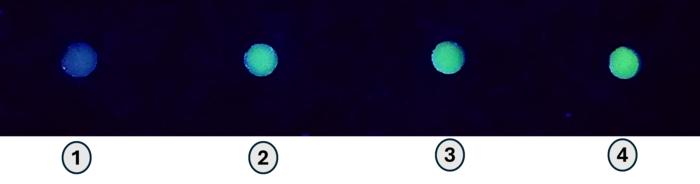
For real-world applicability, the system was adapted into a paper-based format by immobilizing the terbium-gel matrix onto paper discs. This innovation allows the sensor to be easily handled, stored, and deployed without elaborate laboratory infrastructure. Samples containing β-glucuronidase treated with the masked 2,3-DHN are applied onto the paper sensor. Subsequent exposure to UV light reveals a pronounced green luminescent signal proportional to enzyme concentration. This visual “turn-on” response is both striking and quantifiable, representing a significant advancement over complex instrumentation typically required for such assays.
One of the most compelling aspects of this technology is its capacity for straightforward analysis. The enhanced luminescence can be detected using a standard UV lamp, and image analysis software such as ImageJ — an open-source and freely available tool — can quantify emission intensity. This approach eliminates the need for expensive fluorescence spectrometers or high-end diagnostic devices, democratizing access to important biomarker detection. The sensor exhibits a limit of detection (LOD) of approximately 185 ng/mL for β-glucuronidase, a remarkable threshold nearing clinical relevance.
To contextualize this sensitivity, β-glucuronidase concentrations in biological fluids exceeding around 1,000 ng/mL are often indicative of severe liver conditions, including decompensated cirrhosis, a common precursor to liver cancer. Detecting enzyme levels well below this pathological range enables early intervention opportunities, potentially improving patient outcomes through timely diagnosis. Moreover, the sensor’s responsiveness to a broad spectrum of related diseases could extend its utility beyond oncology, encompassing neonatal jaundice diagnostics and monitoring drug-induced toxicities.
Before this innovation can enter clinical practice, further validation through extensive clinical trials is essential. The research team acknowledges this need and remains optimistic about the sensor’s translational potential. Efforts will likely focus on evaluating sensor performance across diverse patient populations, investigating long-term stability, and determining compatibility with complex biological samples such as blood or urine. Should these studies affirm initial findings, the terbium-based sensor could significantly reduce the cost and complexity of liver cancer biomarker detection.
This research also underscores the expanding utility of rare earth luminescent materials in biomedical applications. Terbium’s unique photophysical features, including narrow emission bands and prolonged excited state lifetimes, are harnessed here to strike a balance between sensitivity and operational simplicity. The researchers’ novel approach marries inorganic chemistry, materials science, and enzymology, exemplifying interdisciplinary innovation aimed at solving pressing healthcare challenges.
In summary, the terbium-based paper sensor developed at IISc represents a paradigm shift in enzyme detection and cancer biomarker diagnostics. Its clever design exploits enzymatic specificity and photophysical synergy to produce a highly sensitive but user-friendly assay. By enabling rapid and reliable detection of β-glucuronidase without the need for costly instrumentation, this technology holds promise to democratize early cancer detection, ultimately saving lives through early diagnosis and improved disease management.
Subject of Research: Detection of liver cancer biomarker β-glucuronidase using a terbium-based luminescent sensor.
Article Title: Turn-On Luminescent Detection of Liver Cancer Biomarker β-Glucuronidase Using a Terbium-Based Paper Sensor
News Publication Date: 10-Jun-2025
Web References:
https://aces.onlinelibrary.wiley.com/doi/10.1002/asia.202401975
http://dx.doi.org/10.1002/asia.202401975
Image Credits: UM Group
Keywords: β-glucuronidase detection, liver cancer biomarker, terbium luminescence, paper-based sensor, rare earth metals, enzyme assay, fluorescence energy transfer, point-of-care diagnostics, bioluminescent probe, low-cost cancer screening, 2,3-Dihydroxynaphthalene, gel matrix
Tags: biochemistry of β-glucuronidasecost-effective cancer screeningearly cancer diagnostics innovationenzymatic activity in cancerenzyme detection methodologiesIndian Institute of Science researchliver cancer detection technologynovel diagnostic methods for liver cancerphotophysical properties of terbiumresource-limited healthcare solutionsterbium-based luminescent sensorβ-glucuronidase biomarker
