
A recent case report highlighted in the journal “Oncoscience” draws attention to a rare condition known as diffuse cystic adenomyosis, which can closely mimic invasive endometrial cancer on imaging studies. This diagnostic challenge was evident in the case of an 81-year-old female patient who presented with alarming symptoms, including postmenopausal bleeding and an enlarged uterus. The alarming similarity between her condition and aggressive uterine cancer posed a significant dilemma for her healthcare providers, prompting a necessity for more nuanced diagnostic measures.
Adenomyosis is a complex condition characterized by the presence of endometrial tissue within the myometrium, or muscular wall of the uterus. This presence can result in various symptoms, including heavy menstrual bleeding and severe pelvic pain; however, its manifestation in older, postmenopausal women is exceedingly rare. The complexity of diagnosing adenomyosis increases substantially in older demographics, where the typical presentation may be incorrectly classified as a malignant growth. The incident raises important questions about our current methodologies in imaging and diagnosis within gynecology.
In this particular case, standard imaging techniques, such as transvaginal ultrasound and contrast-enhanced Magnetic Resonance Imaging (MRI), indicated possible aggressive uterine cancer. Clinicians, leveraging these diagnostic tools, faced an immediate concern regarding the potential risk of malignancy, subsequently opting for a surgical intervention to preemptively address the perceived threats. This highlights the inherent limitations in imaging methodologies that often result in confusing benign conditions with life-threatening diseases, resulting in substantial anxiety for patients and reliance on surgical procedures that may not be necessary.
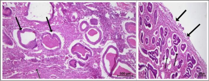
Further validation came from a preoperative biopsy, which revealed endometrial hyperplasia and thickening of the uterine layer, yet no malignancy was detected. This situation underscores the potential for misdiagnosis, particularly in postmenopausal women who may exhibit symptoms that overlap significantly with those of malignancies. In this case, the lack of malignancy discovered post-surgery was a pivotal moment, as subsequent analyses confirmed the presence of a rare form of adenomyosis, particularly the extensive glandular variant, which had been misinterpreted throughout the diagnostic process.
Moreover, the episode calls into question the adequacy of current imaging protocols and diagnostic criteria. The report posits that although MRI is commonly recommended for detecting adenomyosis, the mischaracterization of this condition as similar to invasive cancer represents a significant gap in our diagnostic accuracy that must be addressed. The unique nature of this patient’s symptoms necessitates that gynecologists and radiologists remain vigilant in considering adenomyosis as a differential diagnosis, even in older cohorts.
Advanced diagnostic modalities may provide a way forward in distinguishing between adenomyosis and malignancies. The researchers advocate for using innovative techniques such as Magnetic Resonance Spectroscopy alongside the current imaging criteria in order to delineate benign from malignant conditions more precisely. This isn’t merely an academic exercise; improving diagnostic accuracy can directly alleviate patient distress and reduce the frequency of unnecessary surgical procedures that can carry their own risks.
Given the case, it is crucial to further develop diagnostic frameworks that recognize the full spectrum of adenomyosis, especially in patients presenting with atypical presentations. By refining our approaches, clinicians can help avert unnecessary surgical interventions, which often lead to longer recovery times and emotional distress for patients. It is imperative for the clinical community to acquire a deeper understanding of adenomyosis and to recognize the potential of its presentations in postmenopausal women.
Postmenopausal practitioners and radiologists must remain educated about the nuances of adenomyosis and its possible connections to other conditions. By prioritizing awareness about this rare presentation, medical professionals may be able to revolutionize how they approach screening and diagnostics for suspected cases of uterine disorders. In light of recent findings, there is a clear need for enhanced communication among care teams and increased refinement of imaging and diagnostic standards.
Moving forward, it is imperative to conduct further studies assessing risk factors that may lead postmenopausal patients toward developing adenomyosis. Identifying those at greater risk could inform clinical decision-making and guide the development of more focused research agendas. Establishing a basis for distinguishing the benign forms of adenomyosis from potentially life-threatening conditions affirms a responsible way to advance both research and patient care protocols.
The curious interplay between benign conditions and malignant presentations highlights the essential need for ongoing training and education within medical faculties. Empowering future healthcare professionals with stringent diagnostic skills can mitigate the dangers of misdiagnosis and facilitate a smoother, more effective patient diagnosis journey. Only by elevating our understanding and diagnostic precision can we hope to provide comprehensive care for patients enduring complex gynecological conditions, particularly in their later years of life.
In conclusion, this notable case of diffuse cystic adenomyosis emphasizes the necessity for urgent advancements in the realm of gynecological imaging and diagnostics. By acknowledging the perplexities presented by this and similar conditions, the medical community stands to not only enhance patient care but also uplift the standard of practice relating to women’s reproductive health.
Subject of Research: People
Article Title: Diffuse cystic adenomyosis simulating invasive uterine neoplasm on imaging: A postmenopausal diagnostic perplexity!
News Publication Date: February 10, 2025
Web References: Oncoscience
References: DOI: 10.18632/oncoscience.615
Image Credits: Copyright: © 2025 Devalla et al.
Keywords: adenomyosis, invasive uterine neoplasm, MRI, diagnostic challenges, postmenopausal women, endometrial hyperplasia, surgical intervention, cancer research.
Tags: adenomyosis in older womendiagnostic dilemmas in healthcarediffuse cystic adenomyosisendometrial cancer mimicryhealthcare provider decision-makingimaging challenges in gynecologymanaging abnormal uterine symptomsMRI in gynecological assessmentsnuanced diagnostic measures for women’s healthpostmenopausal bleeding diagnosisrare uterine conditionstransvaginal ultrasound limitations

