In a groundbreaking development within the realm of biological imaging, researchers from Osaka University have unveiled a highly innovative method of Raman microscopy that promises to revolutionize the way we visualize cellular structures and molecular interactions. The current state of biological imaging often encounters limitations largely due to weak Raman signals, which are typically overwhelmed by background noise, leading to images that lack the clarity desired for intricate biological studies. However, the research team has introduced a technique that not only addresses these challenges but also enhances the overall quality of the imagery produced.
Raman microscopy is acclaimed for its ability to provide detailed chemical information about biological samples, particularly regarding the molecular composition of cells. However, capturing high-resolution images has been a perennial challenge, primarily due to the movement of samples during imaging and the low intensity of the Raman signals produced. The Osaka University team has managed to develop a revolutionary microscope that circumvents these issues by allowing previously frozen samples to be imaged without any motion. This innovation ensures that samples remain stable during the acquisition process, dramatically improving the quality of the final images obtained.
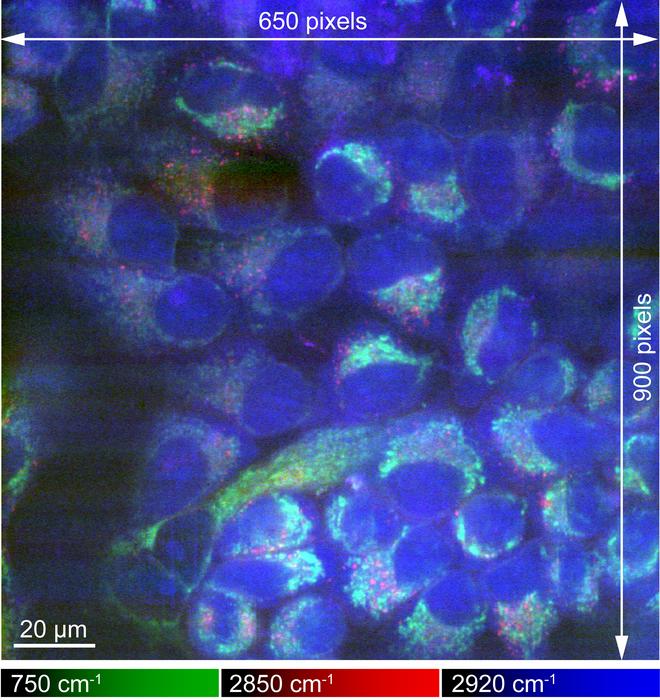
The methodology employed by the researchers centers around maintaining the temperature of biological specimens even as they undergo imaging. By freezing the samples and eliminating potential motion artifacts, the researchers were able to achieve images that are significantly brighter—up to eight times more luminous than earlier iterations of Raman microscopy. Lead author Kenta Mizushima elucidates that this approach resulted in high contrast between the Raman signals and the background noise, thereby leading to greater resolution and expansive fields of view.
A significant advantage of this new technique is its non-invasive nature; it does not require any staining or chemical treatment to secure the cells in place. This allows researchers to capture imagery that is much more representative of the natural state of the cells and the processes occurring within them. Conventional methods often alter the cellular environment and can introduce artifacts that obscure the biological truth.
Moreover, the freezing process employed by the research team appears to successfully conserve the physicochemical states of various proteins within the cells. This is a critical breakthrough since traditional chemical fixing methods can compromise structural integrity, and the lack of such interference ensures a more accurate portrayal of molecular interactions. The implications of this development could extend far beyond imaging, potentially leading to new insights in our understanding of cellular processes, disease mechanisms, and therapeutic interventions.
The versatility of Raman microscopy cannot be understated. As pointed out by senior author Katsumasa Fujita, the great strength of this approach is the complementarity it offers to other imaging techniques. The biological realm is complex—researchers frequently require various types of imaging data to comprehend the whole picture. The ability to collect detailed images while also acquiring insightful molecular data represents a profound advancement in biological sciences, particularly in medical research and pharmaceutical development.
The potential applications of this enhanced imaging technique are vast. It could have profound implications for fields such as histopathology, where precise imaging of tissue samples is crucial for diagnosing diseases, including cancer. Furthermore, its capability to reveal chemical states could allow for more effective tracking of disease progression and the efficacy of therapeutic strategies.
As the team conducted their research, they found ways to refine the imaging process even further. They utilized state-of-the-art cooling technologies to ensure that samples remained at optimal temperatures during imaging, mitigating risks associated with sample degradation. This careful attention to detail ensures that the results produced are of the highest fidelity, allowing researchers to confidently draw conclusions based on their observations.
With the continuous demand for improved imaging modalities in the life sciences, this study could mark a turning point in how researchers engage with biological data. The intricate relationship between molecular structure and function will become increasingly accessible, providing new avenues for exploration in cellular biology. The scientific community has long desired a technique that could offer both high-resolution images and detailed chemical information, and the Osaka University team has surmounted this hurdle.
The ramifications of these findings extend to educational institutions and labs worldwide, where the need for advanced microscopy techniques is urgent. Researchers operating in various fields will likely utilize this new approach to enhance their understanding of cellular dynamics, offering hope for future breakthroughs in both basic and applied sciences.
The transformative power of this research can be felt across multiple domains within the biological sciences. Ara Fuldy, a prominent figure in microscopy research, emphasizes that this methodology could accelerate studies aimed at probing the biochemical pathways active in diseases, thereby directly contributing to the advancement of personalized medicine. The insights garnered from these high-resolution images could facilitate a more pointed approach to targeting therapies and interventions.
In conclusion, the innovative advancements in high-resolution Raman microscopy presented by Osaka University are undoubtedly a significant milestone in the biological imaging landscape. The researchers have opened new channels for understanding molecular interactions and cellular behaviors, potentially leading to breakthroughs in medical diagnostics and therapeutics. As the scientific community calls for more detailed and representative biological data, this work stands to fulfill those needs, pushing the boundaries of what is possible in the field of life sciences.
Subject of Research: High-resolution Raman microscopy of cryofixed biological specimens
Article Title: Raman microscopy of cryofixed biological specimens for high-resolution and high-sensitivity chemical imaging
News Publication Date: 12-Dec-2024
Web References: http://dx.doi.org/10.1126/sciadv.adn0110
References: Available in the publication
Image Credits: Sci. Adv. 10, eadn0110 (2024)
Keywords: Raman microscopy, cryofixing, biological imaging, cellular biology, molecular interaction, advanced microscopy techniques, high-resolution imaging, medical diagnostics, pharmaceutical development.


