Cardiovascular diseases, including stroke and myocardial infarction, are the world’s leading causes of mortality, accounting for over 18 million deaths a year. A team of KIT researchers has now identified a new cell type in blood vessels responsible for vascular growth. This discovery may allow for novel therapeutic strategies to treat ischemic cardiovascular diseases, i.e. diseases that are caused by reduced or absent blood flow. Nature Communications (DOI: 10.1038/s41467-024-47434-x)
Credit: Zoologisches Institut, KIT
Cardiovascular diseases, including stroke and myocardial infarction, are the world’s leading causes of mortality, accounting for over 18 million deaths a year. A team of KIT researchers has now identified a new cell type in blood vessels responsible for vascular growth. This discovery may allow for novel therapeutic strategies to treat ischemic cardiovascular diseases, i.e. diseases that are caused by reduced or absent blood flow. Nature Communications (DOI: 10.1038/s41467-024-47434-x)
In our body, a large network of blood vessels distributes blood across our organs and thereby ensures that the active cells are supplied with sufficient oxygen and nutrients to function and to maintain heart beat and brain activities for example. Occlusion of blood vessels and compromising oxygen delivery may cause neuronal or cardiac cell death culminating in stroke or heart attack. Revascularization , i. e. restoring vascular perfusion and promoting tissue regeneration, requires functional blood vessels, but how to effectively revascularize organs still is an unsolved clinical question.
Since each organ fulfills a different physiological function, vascular branching patterns differ across organs. It has long been a mystery how such unique, so-called organo-typical vascular structures develop.
From a therapeutic point of view, it is believed that understanding the organ-specific molecular control of vascular growth and patterning could open the doors for developing personalized medicine strategies to combat cardiovascular and neurodegenerative diseases and cancer.
Pioneer Cells Move inside the Vessel Walls
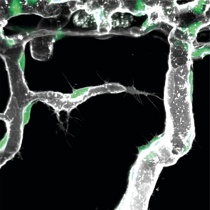
Scientists of the KIT led by Professor Ferdinand le Noble, Head of the Department of Cell and Developmental Biology and Director of the Zoological Institute of KIT, now discovered that one crucial element contributing to organ-dependent variability in vascular branching involves the activation of a novel vascular cell type they coined endothelial L-tip cell or pioneer cell. Pioneer cells reside inside the inner layer that lines the blood vasculature, the so-called endothelium.
Using high-end imaging techniques, the scientists found that pioneer cells move inside the vessel wall. Once they come into contact with specific signals produced by cells in the surrounding organ, pioneer cells start to make new blood vessels. To elucidate which cells produce such signals and how these signals are sensed to promote pioneer cell differentiation, the scientists used a recently developed technique called single cell sequencing.
Molecular Cocktail Encodes the Time and Place of Blood Vessel Formation
Dr. Laetitia Preau, first author of the paper, explains: “Single cell sequencing combines detailed RNA sequencing of individual cells with bio-informatic analyses and allows precise identification of cell subtypes and the molecules these cells produce for cell-to-cell communication. Using this technique, we discovered that the vascular patterning is encoded by a distinct set of molecules that can only be sensed by a subset of endothelial cells to promote vessel growth.”
The cells in the tissue produce an organ-specific set of molecules that encode the instruction how to make a new blood vessel at that particular place and time. Once the prospective pioneer cell senses and unravels this specific tissue-derived molecular code, it will initiate the vascular growth process.
Foundations for New Therapeutic Approaches
It turned out that several organ-specific vascular growth code molecules are drug-targetable, i.e. react to externally added chemicals. Professor Ferdinand le Noble says: “To explore the therapeutic avenues, we are collaborating with chemists, tissue engineers, and artificial intelligence (AI) specialists at the 3ROCKIT platform of the Health Technologies Center established recently at Karlsruhe Institute of Technology (KITHealthTech). We hope to identify novel smart molecules to target the vascular growth process that may benefit patients suffering from ischemic cardiovascular diseases, such myocardial infarction and stroke, as well as from certain forms of cancer.”
The study was financed by the German Research Foundation (DFG) and carried out by KIT in cooperation with the German Center for Cardiovascular Research (DZHK) at its partners sites in Heidelberg and Munich and the Max Planck Institute for Molecular Biomedicine in Münster.
Original Publication:
Laetitia Préau, Anna Lischke, Melanie Merkel, Neslihan Oegel, Maria Weissenbruch, Andria Michael, Hongryeol Park, Dietmar Gradl, Christian Kupatt, Ferdinand le Noble: Parenchymal cues define Vegfa-driven venous angiogenesis by activating a sprouting competent venous endothelial subtype. Nature Communications. DOI: 10.1038/s41467-024-47434-x
https://www.nature.com/articles/s41467-024-47434-x
More Information on KITHealthTech
Journal
Nature Communications
DOI
10.1038/s41467-024-47434-x
Method of Research
Experimental study
Subject of Research
Cells
Article Title
Parenchymal cues define Vegfa-driven venous angiogenesis by activating a sprouting competent venous endothelial subtype
Article Publication Date
10-Apr-2024


