(Memphis, Tenn – September 28, 2023) Many cancers are caused by fusion oncoproteins, molecules that aberrantly form when a rearrangement of DNA results in parts of two different proteins being expressed as one. Several fusion oncoproteins spontaneously form condensates inside cells that promote cancer development. New research by St. Jude Children’s Research Hospital established a method to study this biophysical process in cells, then used that information as a launchpad to predict the behavior of other fusion oncoproteins. The findings, which offer insight into fusion oncoprotein-driven cancers, were published today in Nature Communications.

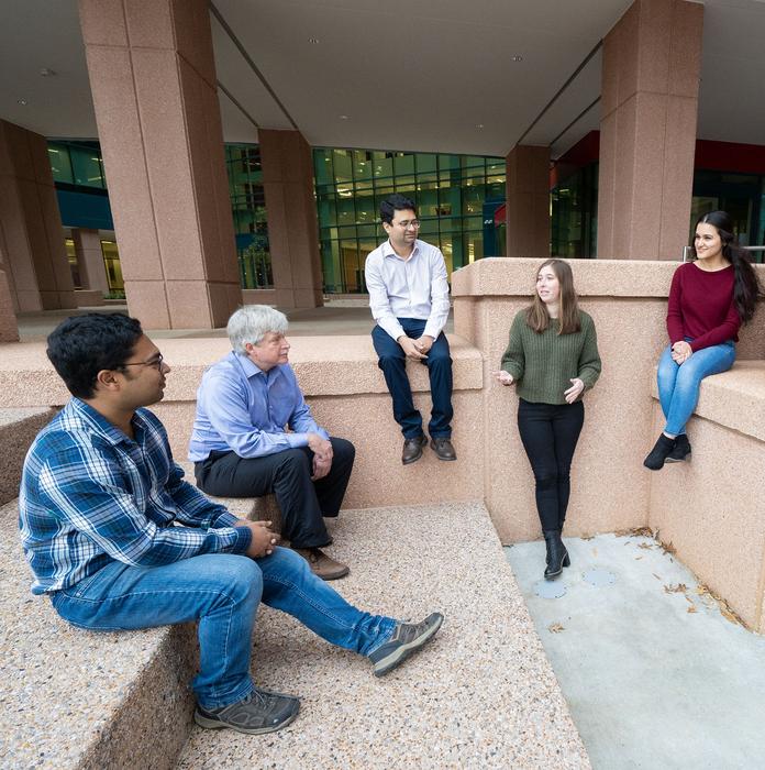
Credit: St. Jude Children’s Research Hospital
(Memphis, Tenn – September 28, 2023) Many cancers are caused by fusion oncoproteins, molecules that aberrantly form when a rearrangement of DNA results in parts of two different proteins being expressed as one. Several fusion oncoproteins spontaneously form condensates inside cells that promote cancer development. New research by St. Jude Children’s Research Hospital established a method to study this biophysical process in cells, then used that information as a launchpad to predict the behavior of other fusion oncoproteins. The findings, which offer insight into fusion oncoprotein-driven cancers, were published today in Nature Communications.
While genes define everything about us, they are not immutable. Genes are made of DNA, which is constantly being read and replicated. Errors can occur, and sometimes a piece of DNA can break and reattach at a different location. This can lead to two previously independent genes being glued together, resulting in a fusion protein. These unnatural proteins retain properties of both original components, which can have disastrous consequences for cells.
“Fusion proteins have been shown to be oncogenic drivers in upwards of 15% of human cancers,” said Richard Kriwacki, Ph.D., St. Jude Department of Structural Biology. These fusion oncoproteins can interfere with cellular regulatory pathways involved in cell growth and differentiation, leading to uncontrolled cell division and cancer.
Secrets in the droplets
“We hypothesized that gaining the ability to form condensates could be linked with the oncogenic properties of fusion oncoproteins,” Kriwacki explained. Biomolecular condensates can form through a process called liquid-liquid phase separation, in which biomolecules separate from the surrounding local environment and form their own compartment, akin to oil droplets in water. Condensates have been shown to be very powerful tools for a cell to regulate many different processes. However, when a fusion oncoprotein has the ability to form a condensate, it can wreak havoc in our cells.
Kriwacki, along with collaborators set out to uncover how interwoven fusion oncoproteins were with the process of phase separation.
The code of fusion oncoprotein condensate behavior
The researchers initially examined 166 fusion oncoproteins in cells to observe if they phase separate. Then they categorized them, which was no small feat, according to co-first author Hazheen Shirnekhi, Ph.D., St. Jude Department of Structural Biology.
“The condensates were all different sizes, different shapes, and located in different areas of the cell,” Shirnekhi said. “It was difficult for any computer program to recognize the condensates in an unbiased manner, so we had to do this manually. It took a lot of time.”
This effort revealed that 58% of the fusion oncoproteins examined formed condensates, opening the door to additional insights.
“We found that a large number of those fusion oncoproteins that form condensates, especially in the nucleus, had functional features associated with regulation of gene expression,” Kriwacki said. “The cytoplasmic fusion oncoproteins forming condensates had functional features associated with regulation of cell signaling.” These observations suggest that the fusion oncoproteins elicit their oncogenic properties by altering gene regulation or cell signaling pathways through formation of condensates.
Machine learning reveals scope of phenomenon
In addition to those links to cellular functions, patterns began to emerge within the protein sequences of the fusion oncoproteins that form condensates. These patterns involve so-called physicochemical features, such as number of polar amino acids, charged groups or disordered regions.
“When we looked at the sequences of the condensate-forming fusion oncoproteins, we noticed features that are distinct from the condensate-negative fusion oncoproteins,” explained co-first author Swarnendu Tripathi, Ph.D., St. Jude Department of Structural Biology. “That motivated us to select 25 non-redundant features and use data science to predict whether a fusion oncoprotein forms condensates or not.”
This data science aspect allowed the researchers to use their 166-sample groundwork to train a machine-learning algorithm using those 25 features. The computational model was then applied to predict the condensate-forming behavior of ~3,000 additional fusion oncoproteins associated with different cancer types.
The model predicted that upwards of 67% of those additional fusion oncoproteins likely form condensates. The condensate-forming predictions were tested for a subset of fusion oncoproteins. “The model was shown to be 80% accurate in independent testing with fusions not used in the training,” Tripathi noted.
This research establishes the foundational framework for determining the mechanisms underlying the oncogenic properties of fusion oncoproteins to enable their targeted inhibition through pharmaceutical agents or alternative approaches. “We’re looking to address the relationship between condensate formation, alteration of gene expression and oncogenesis,” Kriwacki explained. “We’re working with collaborators so that we can address this causality question in as rigorous a way as possible.” As Kriwacki highlighted, “By obtaining a grasp of the underlying mechanisms, we are setting the stage for potential innovative therapeutic approaches against fusion oncoprotein-driven cancers.”
Authors and funding
The study’s other co-first author was Scott Gorman, formerly of St. Jude. The study’s other authors include Bappaditya Chandra, David Baggett, Cheon-Gil Park, Ramiz Somjee, Benjamin Lang, Seyed Mohammad Hadi Hosseini, Brittany Pioso, Ilaria Iacobucci, Qingsong Gao, Michael Edmonson, Stephen Rice, Xin Zhou, John Bollinger, Madan Babu, Charles Mullighan and Jinghui Zhang, of St. Jude; Diana Mitrea and Michael White, formerly of St. Jude, Yongsheng Li and Stephen Yi of the University of Texas at Austin; Daniel McGrail of Cleveland Clinic; Daniel Jarosz of Stanford University School of Medicine; and Nidhi Sahni of the University of Texas MD Anderson Cancer Center and Baylor College of Medicine.
The study was supported by grants from the National Institutes of Health (R35 GM137836, R35 GM133658), Komen Foundation grants (CCR19609287, PDF17483544), the National Cancer Institute (P30 CA021765, R35 CA197695, R01 CA246125, U54 CA243124, R01 CA216391, T32 CA236748, K99 CA240689), the National Institute of General Medical Sciences (F32 GM143847), a St. Jude Children’s Research Hospital Chromatin Collaborative award, a Neoma Boadway Fellowship from St. Jude Children’s Research Hospital, the Cancer Prevention and Research Institute of Texas (RR160021, RP220292), a SummerPlus Program Fellowship from Rhodes College and ALSAC, the fundraising and awareness organization of St. Jude.
St. Jude Media Relations Contacts
Chelsea Bryant
Desk: (901) 595-0564
Cell: (256) 244-2048
[email protected]
[email protected]
Rae Lyn Rushing
Desk: (901) 595-4419
Cell: (901) 686-2597
St. Jude Children’s Research Hospital
St. Jude Children’s Research Hospital is leading the way the world understands, treats and cures childhood cancer, sickle cell disease and other life-threatening disorders. It is the only National Cancer Institute-designated Comprehensive Cancer Center devoted solely to children. Treatments developed at St. Jude have helped push the overall childhood cancer survival rate from 20% to 80% since the hospital opened more than 60 years ago. St. Jude shares the breakthroughs it makes to help doctors and researchers at local hospitals and cancer centers around the world improve the quality of treatment and care for even more children. To learn more, visit stjude.org, read St. Jude Progress blog, and follow St. Jude on social media at @stjuderesearch.
Journal
Nature Communications
Article Publication Date
28-Sep-2023


