With increased use of screening CT for early detection of lung cancer, many more cases of suspicious lung nodules are being identified. However, differentiation of benign from malignant lung nodules remains a challenge that can only be addressed by tissue biopsy. For nodules near the external body surface, the standard of clinical care is to perform a core biopsy procedure, during which tissue cores are obtained by inserting a biopsy needle into the nodule, and the extracted tissue is evaluated by a highly trained pathologist. Guidance of the biopsy needle by interoperative CT or ultrasound is helpful, but the miss rate on collecting appropriate tissue is high. This results in repeat biopsy procedures that can lead to increased risk of complications. Publishing their work in the Journal of Optical Microsystems, a group of investigators at the University of Arizona have developed a multispectral confocal endomicroscope (MSCE) with a sub-millimeter diameter probe that can fit through the lumen of a 20-gauge introducer needle to image the tissue as the distal tip of the needle. This allows evaluation of lung tissue at the site prior to its extraction by needle biopsy.
Credit: The Authors doi: 10.1117/1.JOM.3.1.011002.
With increased use of screening CT for early detection of lung cancer, many more cases of suspicious lung nodules are being identified. However, differentiation of benign from malignant lung nodules remains a challenge that can only be addressed by tissue biopsy. For nodules near the external body surface, the standard of clinical care is to perform a core biopsy procedure, during which tissue cores are obtained by inserting a biopsy needle into the nodule, and the extracted tissue is evaluated by a highly trained pathologist. Guidance of the biopsy needle by interoperative CT or ultrasound is helpful, but the miss rate on collecting appropriate tissue is high. This results in repeat biopsy procedures that can lead to increased risk of complications. Publishing their work in the Journal of Optical Microsystems, a group of investigators at the University of Arizona have developed a multispectral confocal endomicroscope (MSCE) with a sub-millimeter diameter probe that can fit through the lumen of a 20-gauge introducer needle to image the tissue as the distal tip of the needle. This allows evaluation of lung tissue at the site prior to its extraction by needle biopsy.
“The hope is that this added imaging capability will provide the physician with a better view of the tissue to be collected and lead to fewer complications,” said Arthur Gmitro of the University of Arizona, one of the researchers.
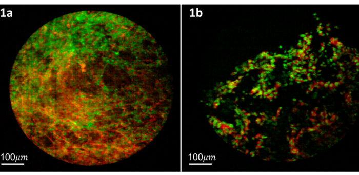
To accomplish this goal, the researchers developed a multispectral fluorescence line-scan confocal endomicroscope with 20 spectral channels that operates at real-time frame rates up to 10 full multispectral frames per second. The choice of 20 spectral channels across the fluorescence emission range of 500 – 750 nm allows multiple fluorescent dyes with overlapping emission spectra to be simultaneously imaged and uniquely identified via spectral unmixing. The system employs a fiber optic imaging bundle with 30,000 elements that works as a tissue contact probe with a field of view of 0.75 mm.
Demonstration experiments were carried out using a combination of two FDA-approved fluorescent dyes, proflavine, which stains the cell nucleus, and rose bengal, which stains proteinaceous connective tissue. Imaging was done in the lung of euthanized Sprague Dawley rats under protocols approved by Institutional Animal Care and Use Committee. The rat experiments were done with the fiber probe inserted through the introducer needle into the lung and topical administration of the proflavine/rose-bengal dye mixture at the distal tissue site. The rat experiments provided a model for how this system would be used in the clinical setting and what normal lung tissue looked like with the MSCE imaging system. Under institutional review board-approved protocols, imaging was also done on fresh tissue samples obtained from patients undergoing conventional core biopsy procedures. Core biopsy tissue from consenting patients was stained by application of the dye mixture directly to the tissue, followed shortly afterward by a quick rinse and then imaging with the MSCE imaging system. The human lung tissue imaging allowed evaluation of what various pathologic conditions (lung cancer, inflammation, and necrosis) look like.
Read the Gold Open Access paper by Li et al., “Multispectral confocal endomicroscopy in lung biopsy guidance,” J. Opt. Microsystems 3(1) 011002 (2023), doi: 10.1117/1.JOM.3.1.011002.
Journal
Journal of Optical Microsystems
DOI
10.1117/1.JOM.3.1.011002
Article Title
Multispectral confocal endomicroscopy in lung biopsy guidance
Article Publication Date
4-Jan-2023


