Over the last two decades, microscopy has seen unprecedented advances in speed and resolution. However, cellular structures are essentially three-dimensional, and conventional super-resolution techniques often lack the necessary resolution in all three directions to capture details at a nanometer scale. A research team led by Göttingen University, including the University of Würzburg and the Center for Cancer Research in the US, investigated a super-resolution imaging technique that involves combining the advantages of two different methods to achieve the same resolution in all three dimensions; this is “isotropic” resolution. The results were published in Science Advances.
Credit: Alexey Chizhik
Over the last two decades, microscopy has seen unprecedented advances in speed and resolution. However, cellular structures are essentially three-dimensional, and conventional super-resolution techniques often lack the necessary resolution in all three directions to capture details at a nanometer scale. A research team led by Göttingen University, including the University of Würzburg and the Center for Cancer Research in the US, investigated a super-resolution imaging technique that involves combining the advantages of two different methods to achieve the same resolution in all three dimensions; this is “isotropic” resolution. The results were published in Science Advances.
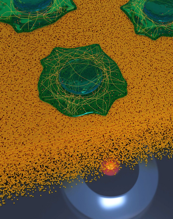
Despite tremendous improvements in microscopy, there still exists a remarkable gap between resolution in all three dimensions. One of the methods that can close this gap and achieves a resolution in the nanometer range is metal-induced energy transfer (MIET) imaging. The exceptional depth resolution of MIET imaging was combined with the extraordinary lateral resolution of single-molecule localization microscopy, in particular with a method called direct stochastic optical reconstruction microscopy (dSTORM). The novel technique based on this combination allows researchers to achieve isotropic three-dimensional super-resolution imaging of sub-cellular structures. In addition, the researchers implement dual-color MIET-dSTORM enabling them to image two different cellular structures in three dimensions, for example microtubules and clathrin coated pits – tiny structures within cells – that exist together in the same area.
“By combining the established concepts, we developed a new technique for super-resolution microscopy. Its main advantage is it enables extremely high resolution in three dimensions, despite using a relatively simple setup,” says Dr Jan Christoph Thiele, first author of the publication, Göttingen University. “This will be a powerful tool with numerous applications to resolve protein complexes and small organelles with sub-nanometer accuracy. Everyone who has access to confocal microscope technology with a fast laser scanner and fluorescence lifetime measurements capabilities should try this technique,” says Dr Oleksii Nevskyi, one of the corresponding authors.
“The beauty of the technique is its simplicity. This means that researchers around the world will be able to implement the technology into their microscopes quickly,” adds Professor Jörg Enderlein who led the research team at the Biophysics Institute, Göttingen University. This method shows promise to become a powerful tool for multiplexed 3D super-resolution microscopy with extraordinary high resolution and a variety of applications in structural biology.
Original publication: Thiele et al, Isotropic three-dimensional dual-color super-resolution microscopy with metal-induced energy transfer, Science Advances 2022. DOI: 10.1126/sciadv.abo2506
Contact:
Professor Jörg Enderlein
University of Göttingen
Third Institute of Physics – Biophysics
Friedrich-Hund-Platz 1, 37077 Göttingen
Tel: +49 551 39 26908
Email: [email protected]
https://www.joerg-enderlein.de/
Dr Oleksii Nevskyi
University of Göttingen
Third Institute of Physics – Biophysics
Friedrich-Hund-Platz 1
37077 Göttingen
Tel.: +49 551 39 22297
Email: [email protected]
https://www.joerg-enderlein.de/galerie
Dr Jan Christoph Thiele
University of Göttingen
Third Institute of Physics – Biophysics
Friedrich-Hund-Platz 1
37077 Göttingen
Tel.: +49 551 39 26909
Email: [email protected]
https://www.joerg-enderlein.de/galerie
Journal
Science Advances
Method of Research
Imaging analysis
Subject of Research
Not applicable
Article Title
Isotropic three-dimensional dual-color super-resolution microscopy with metal-induced energy transfer
Article Publication Date
8-Jun-2022