• HOME
  • NEWS
  • EXPLORE
    • CAREER
      • Companies
      • Jobs
    • EVENTS
    • iGEM
      • News
      • Team
    • PHOTOS
    • VIDEO
    • WIKI
  • BLOG
  • COMMUNITY
    • FACEBOOK
    • INSTAGRAM
    • TWITTER
Sunday, May 3, 2026
BIOENGINEER.ORG
No Result
View All Result
  • Login
  • HOME
  • NEWS
  • EXPLORE
    • CAREER
      • Companies
      • Jobs
        • Lecturer
        • PhD Studentship
        • Postdoc
        • Research Assistant
    • EVENTS
    • iGEM
      • News
      • Team
    • PHOTOS
    • VIDEO
    • WIKI
  • BLOG
  • COMMUNITY
    • FACEBOOK
    • INSTAGRAM
    • TWITTER
No Result
View All Result
Bioengineer.org
No Result
View All Result
Home NEWS Science News Biology

Method offers sharper images of lung disease in living mice

Bioengineer by Bioengineer
March 9, 2022
in Biology
Reading Time: 2 mins read
0
3D image of mouse lungs
Share on FacebookShare on TwitterShare on LinkedinShare on RedditShare on Telegram

In a first for X-ray imaging, researchers in Sweden generated high resolution, three-dimensional images of the lungs of free-breathing mice, without using mechanical ventilation. The technique developed at KTH Royal Institute of Technology could enable research on lung disease to be performed without risking injury to mice through artificial respiration.

3D image of mouse lungs

Credit: KTH Royal Institute of Technology

In a first for X-ray imaging, researchers in Sweden generated high resolution, three-dimensional images of the lungs of free-breathing mice, without using mechanical ventilation. The technique developed at KTH Royal Institute of Technology could enable research on lung disease to be performed without risking injury to mice through artificial respiration.

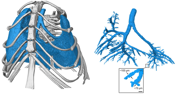
Reporting in Communications Physics, the researchers showed that propagation-based phase-contrast X-ray tomography can be used to study the tracheobronchial tree down to the terminal bronchioles – which are as thin as strands of human hair – in free-breathing mice. What’s more, they were able to do it with low radiation doses, which allows for repeated imaging without harm to the animals.

Mice are typically used in lung disease research due to the similarity of their respiratory system to that of humans. However, the animals need to be placed on ventilators that control their breathing, in order to avoid blurry images.

“Our work shows that it is possible to completely avoid using ventilators, to let the animals breathe themselves, and still get very sharp X-ray images in both 2D and 3D,” says KTH researcher Kian Shaker.

Non-invasive imaging of lung diseases in animals will enable a greater understanding of similar conditions in humans, Shaker says. The images are relevant because they visualize even the thinnest bronchial branches, measuring less than 0.1 mm in diameter in mice and which end in clusters of alveoli, he says.

The footprint for the imaging set up is also smaller than what is required for synchrotron facilities where high-resolution respiratory imaging of animals is typically performed.

In the future, these images indicate what Shaker and his colleagues hope will be possible also for imaging human lungs, where the smallest airways have diameters below 2 mm and are invisible with the technology used in hospitals today.

The research was funded by the Knut and Alice Wallenberg Foundation.


Journal

Communications Physics

DOI

10.1038/s42005-021-00760-8

Method of Research

Experimental study

Article Title

Phase-contrast X-ray tomography resolves the terminal bronchioles in free-breathing mice

Article Publication Date

9-Dec-2021

Share12Tweet8Share2ShareShareShare2

Related Posts

Successful Birth Following Uterus Transplant Marks Medical Breakthrough — Biology

Successful Birth Following Uterus Transplant Marks Medical Breakthrough

May 1, 2026
Cockatoos Mimic Peers to Sharpen Adaptation Skills, Study Finds — Biology

Cockatoos Mimic Peers to Sharpen Adaptation Skills, Study Finds

May 1, 2026

Gut Microbe’s Sulfated Bile Acid Eases Pediatric Sepsis

May 1, 2026

AI Breakthrough Solves One of Science’s Most Challenging Math Problems

May 1, 2026

POPULAR NEWS

  • Research Indicates Potential Connection Between Prenatal Medication Exposure and Elevated Autism Risk

    833 shares
    Share 333 Tweet 208
  • New Study Reveals Plants Can Detect the Sound of Rain

    716 shares
    Share 286 Tweet 179
  • Scientists Investigate Possible Connection Between COVID-19 and Increased Lung Cancer Risk

    67 shares
    Share 27 Tweet 17
  • Salmonella Haem Blocks Macrophages, Boosts Infection

    61 shares
    Share 24 Tweet 15

About

We bring you the latest biotechnology news from best research centers and universities around the world. Check our website.

Follow us

Recent News

AI Boosts Cost-Effectiveness in UK Breast Screening

Shock-Scattering Micro-Histotripsy Boosts Fine Needle Biopsies

Simulating Neurodevelopment Care for Children with Heart Disease

Subscribe to Blog via Email

Enter your email address to subscribe to this blog and receive notifications of new posts by email.

Join 82 other subscribers
  • Contact Us

Bioengineer.org © Copyright 2023 All Rights Reserved.

Welcome Back!

Login to your account below

Forgotten Password?

Retrieve your password

Please enter your username or email address to reset your password.

Log In
No Result
View All Result
  • Homepages
    • Home Page 1
    • Home Page 2
  • News
  • National
  • Business
  • Health
  • Lifestyle
  • Science

Bioengineer.org © Copyright 2023 All Rights Reserved.