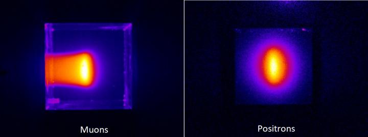
The quality of muon beams can now be assessed thanks to a new technique that has produced the first known images of these high-energy particles.

Credit: Seiichi Yamamoto
A new technique has taken the first images of muon particle beams. Nagoya University scientists designed the imaging technique with colleagues in Osaka University and KEK, Japan and describe it in the journal Scientific Reports. They plan to use it to assess the quality of these beams, which are being used more and more in advanced imaging applications.
Muons are charged particles that are 207 times the mass of electrons. They naturally form when cosmic rays strike atoms in the upper atmosphere, showering down onto every part of Earth’s surface. They can penetrate through hundreds of meters of solids before being absorbed.
Scientists have used naturally occurring muon particles as a way to peek through huge solid structures. For example, in 2017 scientists announced they had found a hidden chamber inside the Khufu Pyramid of Giza by comparing the muon intensities measured by detectors located inside and outside the pyramid. Particle accelerator facilities can now also generate muon beams, which are used in a variety of applications, like non-destructive X-ray fluorescence spectroscopy. Muon beams are also expected to be adapted for cancer radiotherapy.
Nagoya University biomedical nuclear scientist Seiichi Yamamoto and colleagues developed a new imaging technique that they say shows promise for quality assessment and research of muon beams, and should be of benefit for muon radiotherapy in the future.
The technique depends on a phenomenon that occurs when charged particles travel through transparent media, like water. Water slows light down relative to high-energy particles. Particles moving faster than light cause something similar to the sonic boom we hear when a jet plane breaks through the sound barrier. In the case of the particles, an ‘optical boom’, called the Cherenkov effect, causes a brief flash.
Yamamoto and his colleagues imaged this effect with a special camera when a muon beam was directed through water or a plastic scintillator block. The technique allowed them to image muons and the positrons that form when muons decay. This helped them measure the beam’s range through the water or plastic scintillator, and the deviation of its momentum, as well as clarify the direction of positron movement.
“The system is compact, low cost and easy to use, showing promise as a tool for quality assessment in muon beam facilities,” says Yamamoto.
###
The study, “Optical imaging of muons,” was published in the journal Scientific Reports on November 26, 2020, at DOI:10.1038/s41598-020-76652-8.
About Nagoya University, Japan
Nagoya University has a history of about 150 years, with its roots in a temporary medical school and hospital established in 1871, and was formally instituted as the last Imperial University of Japan in 1939. Although modest in size compared to the largest universities in Japan, Nagoya University has been pursuing excellence since its founding. Six of the 18 Japanese Nobel Prize-winners since 2000 did all or part of their Nobel Prize-winning work at Nagoya University: four in Physics – Toshihide Maskawa and Makoto Kobayashi in 2008, and Isamu Akasaki and Hiroshi Amano in 2014; and two in Chemistry – Ryoji Noyori in 2001 and Osamu Shimomura in 2008. In mathematics, Shigefumi Mori did his Fields Medal-winning work at the University. A number of other important discoveries have also been made at the University, including the Okazaki DNA Fragments by Reiji and Tsuneko Okazaki in the 1960s; and depletion forces by Sho Asakura and Fumio Oosawa in 1954.
Website: http://en.
Media Contact
Seiichi Yamamoto
[email protected]
Original Source
http://en.
Related Journal Article
http://dx.


