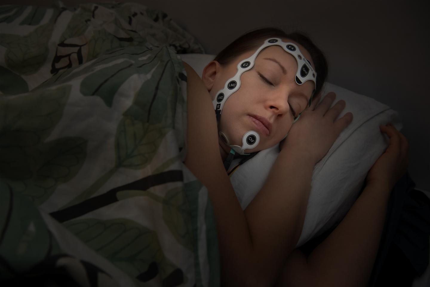
Credit: UEF / Raija Törrönen
A new neural network developed by researchers at the University of Eastern Finland and Kuopio University Hospital enables an easy and accurate assessment of sleep apnoea severity in patients with cerebrovascular disease. The assessment is automated and based on a simple nocturnal pulse oximetry, making it possible to easily screen for sleep apnoea in stroke units.
Up to 90% of patients experiencing a stroke have sleep apnoea, according to earlier studies conducted at Kuopio University Hospital. If left untreated, sleep apnoea can reduce the quality of life and rehabilitation of patients with stroke and increase the risk for recurrent cerebrovascular events.
“Although screening of sleep apnoea is recommended for patients with cerebrovascular disease, it is rarely done in stroke units due to complicated measurement devices, time-consuming manual analysis, and high costs,” Researcher Akseli Leino from the University of Eastern Finland says.
In the new study, researchers developed a neural network to assess the severity of sleep apnoea in patients with acute stroke and transient ischaemic attack (TIA) by using a simple nocturnal oxygen saturation signal. The apnoea-hypopnea index, which represents the number of apnoea and hypopnea events per hour, is commonly used in the diagnostics of sleep apnoea. When the researchers compared the results of manual scoring and those obtained using the new neural network, the median difference was only 1.45 events per hour. The neural network was also 78% accurate in classifying patients into four different categories on the basis of sleep apnoea severity (no sleep apnoea, mild, moderate, severe). The neural network was able to identify moderate and severe sleep apnoea, both of which require treatment, in patients with acute stroke or TIA with a 96% specificity and a 92% sensitivity.
“The neural network developed in the study enables an easy and cost-effective screening of sleep apnoea in patients with cerebrovascular disease in hospital wards and stroke units. The nocturnal oxygen saturation signal can be recorded with a simple finger pulse oximetry measurement, with no time-consuming manual analysis required,” Medical Physicist Katja Myllymaa from Kuopio University Hospital points out.
###
The study was conducted in collaboration between the Department of Clinical Neurophysiology and the Department of Neurology at Kuopio University Hospital, and the Department of Applied Physics at the University of Eastern Finland. The study was funded by the Academy of Finland, Business Finland, Kuopio University Hospital, the Finnish Cultural Foundation, Kuopio Area Respiratory Foundation, the Research Foundation of the Pulmonary Diseases, the Finnish Anti-Tuberculosis Association Foundation, Päivikki & Sakari Sohlberg Foundation, Paulo Foundation, and Tampere Tuberculosis Foundation.
For further information, please contact:
Early Stage Researcher Akseli Leino, MSc (Tech), [email protected]
Docent, Medical Physicist Katja Myllymaa, PhD, [email protected]
Research article:
Leino A, Nikkonen S, Kainulainen S, Korkalainen H, Töyräs J, Myllymaa S, Leppänen T, Ylä-Herttuala S, Westeren-Punnonen S, Muraja-Murro A, Jäkälä P, Mervaala E, Myllymaa K. Neural network analysis of nocturnal SpO2 signal enables easy screening of sleep apnea in patients with acute cerebrovascular disease. Sleep Med 2020;79. https:/
Media Contact
Akseli Leino
[email protected]
Related Journal Article
http://dx.


